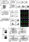
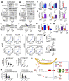
. 2021 Apr;28(4):1174-1192.
doi: 10.1038/s41418-020-00641-7. Epub 2020 Oct 27.

Inhibition of Drp1 SUMOylation by ALR protects the liver from ischemia-reperfusion injury

Affiliations

Inhibition of Drp1 SUMOylation by ALR protects the liver from ischemia-reperfusion injury

Jing Huang et al. Cell Death Differ. 2021 Apr.

Abstract

Hepatic ischemic reperfusion injury (IRI) is a common complication of liver surgery. Although an imbalance between mitochondrial fission and fusion has been identified as the cause of IRI, the detailed mechanism remains unclear. Augmenter of liver regeneration (ALR) was reported to prevent mitochondrial fission by inhibiting dynamin-related protein 1 (Drp1) phosphorylation, contributing partially to its liver protection. Apart from phosphorylation, Drp1 activity is also regulated by small ubiquitin-like modification (SUMOylation), which accelerates mitochondrial fission. This study aimed to investigate whether ALR-mediated protection from hepatic IRI might be associated with an effect on Drp1 SUMOylation. Liver tissues were harvested from both humans and from heterozygous ALR knockout mice, which underwent IRI. The SUMOylation and phosphorylation of Drp1 and their modulation by ALR were investigated. Hepatic Drp1 SUMOylation was significantly increased in human transplanted livers and IRI-livers of mice. ALR-transfection significantly decreased Drp1 SUMOylation, attenuated the IRI-induced mitochondrial fission and preserved mitochondrial stability and function. This study showed that the binding of transcription factor Yin Yang-1 (YY1) to its downstream target gene UBA2, a subunit of SUMO-E1 enzyme heterodimer, was critical to control Drp1 SUMOylation. By interacting with YY1, ALR inhibits its nuclear import and dramatically decreases the transcriptional level of UBA2. Consequently, mitochondrial fission was significantly reduced, and mitochondrial function was maintained. This study showed that the regulation of Drp1 SUMOylation by ALR protects mitochondria from fission, rescuing hepatocytes from IRI-induced apoptosis. These new findings provide a potential target for clinical intervention to reduce the effects of IRI during hepatic surgery.


Conflict of interest statement

The authors declare that they have no conflict of interest.

Figures
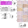
Fig. 1
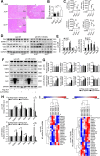
Fig. 1. SUMOylation of Drp1 is increased in transplant livers from patients.
A H&E staining in liver tissues of patients w/i liver transplant (Liver-Tx, n = 5, IRI, 10 h/2 h) or w/o Tx (as control, n = 5). Scale bar, 200 μm, 100 μm. B Liver damage induced by IRI was evaluated by Suzuki’s histological criterion. C Immunohistochemical staining and D western blotting of indicated proteins in human liver tissues w/i Liver-Tx or w/o Tx. GAPDH was used as loading control. E The quantifications of indicated protein contents in human liver tissues (n = 3). F SUMOylation of Drp1 was analyzed by immunoprecipitation (IP) in liver of patients w/i Liver-Tx or w/o Tx. Anti-Drp1 immunoprecipitants were analyzed by western blotting with anti-SUMO1 antibody. G The quantifications of Drp1 SUMOylation in human liver tissues (n = 3). P-values were calculated by Student’s t-test. Data represent mean ± S.D. *P < 0.05; **P < 0.01; ***P < 0.001; ****P < 0.0001. Abbreviations: Liver-Tx liver transplant patients, Ctrl control patients, w/o without, w/i with, p-Drp1 phosphorylated-Drp1.
Fig. 2
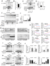
Fig. 2. SUMOylated Drp1 in mitochondria is increased in human transplant livers.
A Western blotting of Drp1 and SUMO1 in cytosolic fraction and mitochondrial compartment from liver tissues of patients w/i Liver-Tx or w/o Tx. GAPDH and COX IV were used as loading controls, respectively. B The quantifications of Drp1 and SUMO1 in cytosolic fraction and mitochondrial compartment from human liver tissues. The proteins in cytosolic fraction and mitochondrial compartment were normalized by GAPDH and COX IV respectively (n = 3). C SUMOylation of Drp1 was analyzed by IP in cytosolic fraction and mitochondrial compartment from liver tissues of patients w/i Liver-Tx or w/o Tx. Anti-Drp1 immunoprecipitants were analyzed by western blotting with anti-SUMO1 antibody. D The quantifications of Drp1 SUMOylation in cytosolic fraction and mitochondrial compartment from human liver tissues (n = 3). E Electron microscopy analysis of liver tissues in patients w/i Liver-Tx or w/o Tx. The arrows in red indicated the elongated mitochondria. Scale bar, 2 μm. F The length of mitochondria was measured in 20 cells of each group, respectively (left). Mitochondrial number (per cell) was counted in 20 cells (middle). Mitochondrial circularity factor in 20 cells (right). The values were recorded from 0 to 1 (0 represents perfect linear, and 1 represents perfect circular). P-values were calculated by Student’s t-test. Data represent mean ± S.D. *P < 0.05; **P < 0.01; ***P < 0.001; ****P < 0.0001. Abbreviations: Liver-Tx liver transplant patients, Ctrl control patients, w/o without, w/i with, Cyto cytosolic fraction, Mito mitochondrial compartment.
Fig. 3
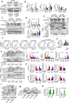
Fig. 3. ALR knockdown increases the expression of SUMO-related proteins after IRI.
A H&E staining in liver tissues of C57BL/6J wild-type mice (ALR+/+, n = 5) and heterozygote knockout of ALR mice w/i (ALR+/−, n = 4) or w/o 2-D08 (24 h, 10 mg/kg) intraperitoneal injection (n = 5) after IRI. Scale bar, 200 μm, 100 μm. B Liver damage induced by IRI was evaluated by Suzuki’s histological criterion. C Liver function parameters (ALT, AST, ALB and LDH) after IRI in serum. D Western blotting of indicated proteins in ALR+/− and ALR+/+ mice w/o or w/i IRI (ALR+/+, n = 4; ALR+/−, n = 4). GAPDH was used as loading control. E The quantifications of indicated proteins in mouse liver (n = 3). F Western blotting of indicated proteins in HepG2 cells transfected with Flag-ALR or ALR-shRNA. Cells were treated either w/i H/R (5 h/2 h) or w/o H/R. GAPDH was used as loading control. G The quantifications of indicated proteins (n = 3). H RT-qPCR of indicated genes in Flag-ALR or ALR-shRNA transfected HepG2 cells. Cells were treated either w/i H/R or w/o H/R (n = 3). I Heatmap of RNA-seq was created in ALR-shRNA and NC-shRNA-transfected HepG2 cells. P-values were calculated by Student’s t-test. Data represent mean ± S.D. *P < 0.05; **P < 0.01; ***P < 0.001. Abbreviations: I/R ischemia-reperfusion, H/R hypoxia/reoxygenation, w/o without, w/i with.
Fig. 4
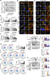
Fig. 4. Insufficiency of ALR gene amplifies Drp1 SUMOylation and recruitment to mitochondria.
A Drp1 SUMOylation was analyzed by IP in liver tissues from ALR+/+ and ALR+/− mice w/o IRI, anti-Drp1 for IP and anti-SUMO1 for western blotting. B The quantifications of Drp1 SUMOylation in mouse livers w/o IRI (n = 3). C Drp1 SUMOylation was analyzed by IP in livers from ALR+/+ and ALR+/− mice after IRI, anti-Drp1 for IP and anti-SUMO1 for western blotting. ALR+/− mice with 2-D08 treatment (24 h, 10 mg/kg) were used as control. D The quantifications of Drp1 SUMOylation in mouse livers w/i IRI (n = 3). E IP of Drp1 SUMOylation in HepG2 cells transfected with Flag-ALR or ALR-shRNA. Cells were either treated w/i H/R or w/o H/R. Anti-Drp1 immunoprecipitants were analyzed by western blotting with anti-SUMO1 antibody. F The quantifications of Drp1 SUMOylation in HepG2 cells (n = 3). G Western blotting of Drp1 and SUMO1 in cytosolic fraction and mitochondrial compartment of HepG2 cells transfected with Flag-ALR (left) or ALR-shRNA (right). Cells were treated under the condition of H/R with prolonged hypoxia time (0–5 h/2 h). GAPDH and COX IV were used as loading controls, respectively. H The quantifications of Drp1 and SUMO1 in cytosolic fraction and mitochondrial compartment of HepG2 cells. The proteins in cytosol and mitochondria were normalized by GAPDH and COX IV, respectively (n = 3). SUMOylation of Drp1 was analyzed by IP in cytosolic fraction and mitochondrial compartment from HepG2 cells transfected either with Flag-ALR (I) or ALR-shRNA (K). Anti-Drp1 immunoprecipitants were analyzed by western blotting with anti-SUMO1 antibody. The quantifications of Drp1 SUMOylation in cytosolic fraction and mitochondrial compartment from Flag-ALR (J) or ALR-shRNA (L) transfected cells (n = 3). P-values were calculated by both Student’s t-test (B, D, F, J, L) and two-way ANOVA test (H). Data represent mean ± S.D. *P < 0.05; **P < 0.01; ***P < 0.001. Abbreviations: I/R ischemia-reperfusion, H/R hypoxia/reoxygenation, w/o without, w/i with, IP immunoprecipitation, IB immunoblotting, Cyto cytosolic fraction, Mito mitochondrial compartment.
Fig. 5
Fig. 5. ALR reciprocally modulates phosphorylation and SUMOylation to restrict Drp1 mitochondrial translocation during hepatic IRI.
A SUMOylation and phosphorylation of Drp1 were analyzed by IP in HepG2 cells transfected with Myc-Drp1 or mutants of S616A, S616D, and 4KR, respectively. Cells were treated w/i or w/o H/R. Anti-Drp1 immunoprecipitants were analyzed by western blotting with anti-SUMO1, anti-p-Drp1 at Ser616 and Ser637 site antibodies. B The quantifications of Drp1 SUMOylation, p-Drp1S616 and p-Drp1S637 in HepG2 cells transfected either with Myc-Drp1 or indicated mutants. The results were normalized to the values of Drp1-WT group (n = 3). C SUMOylation and phosphorylation of Drp1 were analyzed by IP in HepG2 cells transfected with ALR-shRNA. Cells were treated with 2-D08 (18 h, 0 μM, 50 μM, 100 μM). NC-shRNA was used as negative control. Anti-Drp1 immunoprecipitants were analyzed by western blotting with anti-SUMO1, anti-p-Drp1 at Ser616 and Ser637 site antibodies. D The quantifications of Drp1 SUMOylation and p-Drp1S616 in cells with 2-D08 treatment (24 h, 100 μM). The results were normalized to the values of NC-shRNA group without 2-D08 and H/R treatment (n = 3). E IP of Drp1 SUMOylation and western blotting of Drp1, SUMO1 and p-Drp1 in cytosolic fraction and mitochondrial compartment from HepG2 cells transfected with ALR-shRNA. UBA2 was knockdown by siRNA. The β-actin and COX IV were used as loading controls, respectively. F Flow cytometry plots for Annexin V-EGFP and PI staining in HepG2 cells transfected with Flag-ALR or ALR-shRNA under the condition of H/R. The ALR-shRNA cells were transfected with siUBA2 or treated with 2-D08 (24 h, 100 μM). G The percentage of EGFP+/PI+ and EGFP+/PI cells. The values were normalized to negative control group transfected with Flag-tagged vector (n = 3). H Western blotting of indicated proteins in HepG2 cells transfected with ALR-shRNA. The cells were transfected either with Myc-Drp1 WT or 4KR mutant. GAPDH was used as loading control. I The quantifications of indicated proteins were analyzed (n = 3). J Western blotting of p-Drp1S616 and p-Drp1S637 in cytosolic fraction and mitochondrial compartment from ALR-shRNA HepG2 cells transfected either with Myc-Drp1 WT or 4KR mutant. The β-actin and COX IV were used as loading control, respectively. K The quantifications of indicated proteins in cytosolic fraction and mitochondrial compartment were analyzed in cells subject to w/o (upper) or w/i (lower) H/R (n = 3). L IP of Drp1 SUMOylation, western blotting of Drp1, p-Drp1, ATPS-β, NDUFB8, Caspase-3, and cleaved Caspase-3 in HepG2 cells transfected either with Myc-Drp1 WT or 4KR-S616A mutant. GAPDH was used as loading control. M Western blotting of Drp1 in cytosolic fraction and mitochondrial compartment from HepG2 cells transfected either with Myc-Drp1 WT or 4KR-S616A mutant. The β-actin and COX IV were used as loading control, respectively. N The quantifications of Drp1 in cytosolic fraction and mitochondrial compartment (n = 3). P-values were calculated by both Student’s t-test (B, G, I, K, N) and one-way ANOVA (D). Data represent mean ± S.D. *P < 0.05; **P < 0.01; ***P < 0.001. Abbreviations: p-Drp1 phosphorylated-Drp1, H/R hypoxia/reoxygenation, w/o without, w/i with, IP, immunoprecipitation, IB, immunoblotting, Cyto, cytosolic fraction, Mito mitochondrial compartment.
Fig. 6
Fig. 6. ALR blocks Drp1 mitochondrial translocation and maintains the mitochondrial homeostasis after IRI.
A Western blotting of indicated proteins in ALR-shRNA HepG2 cells transfected with siSUMO1, siUBA2, siUbc9, or treated with 2-D08 (18 h, 100 μM) and Ro-3306 (20 h, 35 nM), respectively. GAPDH was used as loading control. B, C Representative images of immunofluorescence staining on Drp1 (green), mitochondria (red), and DAPI (blue) in HepG2 cells with ALR-shRNA-transfected w/o (B) or w/i H/R (C). Scale bar, 7.5, 5, 2 μm. D Quantifications of mitochondrial length in about 50 cells of each group. The results were normalized to the values of ALR-shRNA group. E Quantifications of mitochondrial number (per cell) in about 50 cells. F Quantifications of mitochondrial circularity factor in about 50 cells. The values were recorded as 0 to 1 (0 means perfect linear and 1 means perfect circular). G, H Flow cytometry plots for JC-1 aggregates (red) and monomer (green) in ALR-shRNA HepG2 cells w/o (G) or w/i H/R (H). The cells were treated with siRNA against UBA2 or Ubc9, or treated with 2-D08 (24 h, 100 μM) and Ro-3306 (20 h, 35 nM), respectively. JC-1 ratios for aggregates/monomer were calculated (right) (n = 3). IP of Drp1 SUMOylation, western blotting of Drp1, Mul1, ATPS-β, NDUFB8, Caspase-3 and cleaved Caspase-3 in HepG2 cells transfected either with Flag-ALR (I) or ALR-shRNA (K). The cells were transfected with siMul1. GAPDH was used as loading control. The graphs of indicated ratios in Flag-ALR (J) or ALR-shRNA (L) cells were quantified and normalized to the values of Flag-vector or NC-shRNA transfected with siNC w/o H/R, respectively (n = 3). P-values were calculated by Student’s t-test. Data represent mean ± S.D. *P < 0.05; **P < 0.01; ****P < 0.0001. Abbreviations: H/R hypoxia/reoxygenation, w/o without, w/i with, DAPI 4′,6‐diamidino‐2‐phenylindole.
Fig. 7
Fig. 7. ALR interacts with transcriptional factor YY1 and inhibits UBA2 transcription.
A Co-IP of interaction between ALR and YY1 or YY1AP1 in HepG2 cells transfected with Flag-ALR. Anti-Flag immunoprecipitants were analyzed by western blotting with anti-YY1, anti-YY1AP1 antibodies. Cells were treated w/i or w/o H/R. B Direct interaction between YY1 and ALR was revealed by GST pull-down assays. Input and pull-down samples were both subjected to western blotting with anti-GST and anti-His antibodies. Ten percent of bacterial lysate was used for input. C Western blotting of UBA2, YY1 and YY1AP1 in HepG2 cells transfected with Flag-ALR. Cells were either treated w/i or w/o H/R. GAPDH was used as loading control. D The quantifications of indicated proteins (n = 3). E Western blotting of UBA2, YY1 and YY1AP1 in HepG2 cells transfected with ALR-shRNA. The ALR-shRNA cells were transfected with Flag-ALR for rescue experiment. GAPDH was used as loading control. Cells were either treated w/i or w/o H/R. F The quantifications of indicated proteins (n = 3). G Co-IP of interaction between ALR and YY1 or YY1AP1, western blotting of YY1 and YY1AP1 in cytoplasm and nucleus from HepG2 cells transfected with Flag-ALR. The α-tubulin and lamin A/C were used as loading controls, respectively. H The quantifications of YY1 and YY1AP1 in cytoplasm and nucleus. The proteins were normalized by α-tubulin and lamin A/C, respectively (n = 3). I Representative images of immunofluorescence staining on YY1 (upper)/YY1AP1 (lower) (green), Flag (red) and DAPI (blue) in HepG2 cells transfected with Flag-ALR. Flag-vector was a negative control. Scale bar, 10 μm. J Western blotting of YY1 and YY1AP1 in cytoplasm and nucleus from HepG2 cells transfected with ALR-shRNA. The ALR-shRNA cells were transfected with Flag-ALR for rescue experiment. Cells were either treated w/o or w/i H/R. The α-tubulin and lamin A/C were used as loading controls, respectively. K The quantifications of YY1 and YY1AP1 in cytoplasm and nucleus (n = 3). L EMSA was performed with 5 μl aliquot containing 100 ng of GST-YY1 and 5 pmol of biotin-labeled UBA2 promoter oligonucleotides, YY1 antibody and an unlabeled competitor probe were added as indicated. The unlabeled competitor at a 10- or 50-fold excess was included in the incubation mixture prior to the EMSA. M, N IP of Drp1 SUMOylation, western blotting of UBA2, YY1 or YY1AP1 in HepG2 cells transfected with Flag-ALR. Cells were transfected either with siYY1 (M) or siYY1AP1 (N). Cells were either treated w/i or w/o H/R. GAPDH was used as loading control. O, P IP of Drp1 SUMOylation, western blotting of UBA2, YY1 or YY1AP1 in HepG2 cells transfected with ALR-shRNA. The ALR-shRNA cells were transfected with Flag-ALR for rescue experiment. Cells were transfected with siYY1 (O) or siYY1AP1 (P). Cells were treated w/i or w/o H/R. GAPDH was used as loading control. P-values were calculated by Student’s t-test. Data represent mean ± S.D. *P < 0.05; **P < 0.01; ***P < 0. 001. Abbreviations: H/R hypoxia/reoxygenation, w/o without, w/i with IP immunoprecipitation, IB immunoblotting, DAPI 4′,6‐diamidino‐2‐phenylindole, Mut mutant, Cyto cytoplasm, Nu nucleus.
Fig. 8
Fig. 8. Knockdown of YY1 alleviates mitochondrial dysfunction and apoptosis caused by H/R.
A, B IP of Drp1 SUMOylation, western blotting of YY1, YY1AP1, ATPS-β, NDUFB8, Caspase-3 and cleaved Caspase-3 in HepG2 cells transfected either with Flag-ALR (A) or ALR-shRNA (B) under the condition of H/R. The cells were transfected either with siYY1 or siYY1AP1. GAPDH was used as loading control. C, D The graphs of indicated ratios in Flag-ALR (C) or ALR-shRNA (D) cells were quantified, respectively (n = 3). E, F Western blotting of Drp1 in cytosolic fraction and mitochondrial compartment from Flag-ALR (E) or ALR-shRNA (F) HepG2 cells transfected with siYY1 or siYY1AP1 under the condition of H/R. The β-actin and COX IV were used as loading control, respectively. G, H The quantifications of Drp1 in cytosolic fraction and mitochondrial compartment from Flag-ALR (G) or ALR-shRNA (H) HepG2 (n = 3). IJ Flow cytometry plots for Annexin V-EGFP and PI staining in HepG2 cells transfected with Flag-ALR (I) and ALR-shRNA (J) under the condition of H/R. The cells were transfected with siYY1 or siYY1AP1. K The percentage of EGFP+/PI+ and EGFP+/PI cells. The values were normalized to the negative control groups, respectively (n = 3). L, M The qChIP assays for UBA2 and RPL30 promoter binding using anti-YY1 in HepG2 cells with Flag-ALR (L) or ALR-shRNA (M) transfection. The ALR-shRNA cells were transfected with Flag-ALR for rescue. Cells were treated either w/i or w/o H/R. The results were represented as fold change over control (IgG) (n = 3). N Working model of ALR-maintaining mitochondrial dynamics and preventing apoptosis during IRI. P-values were calculated by Student’s t-test. Data represent mean ± S.D. *P < 0.05; **P < 0.01; ***P < 0.001. Abbreviations: H/R hypoxia/reoxygenation, w/o without, w/i with, IP immunoprecipitation, IB immunoblotting, Cyto cytosolic fraction, Mito mitochondrial compartment.

Similar articles

Cited by

References

    1. Huguet C, Addario-Chieco P, Gavelli A, Arrigo E, Harb J, Clement RR. Technique of hepatic vascular exclusion for extensive liver resection. Am J Surg. 1992;163:602–5. - PubMed
    1. Abdalla EK, Roger N, Jacques B. Hepatic vascular occlusion: which technique? Surg Clin North Am. 2004;84:563–85. - PubMed
    1. Selzner M, Rüdiger HA, Sindram D, Madden J, Clavien PA. Mechanisms of ischemic injury are different in the steatotic and normal rat liver. Hepatology. 2000;32:1280–8. - PubMed
    1. Odeh M. The role of reperfusion-induced injury in the pathogenesis of the crush syndrome. N. Engl J Med. 1991;324:1417–22. - PubMed
    1. Elias-Miró M, Jiménez-Castro MB, Rodés J, Peralta C. Current knowledge on oxidative stress in hepatic ischemia/reperfusion. Free Radic Res. 2013;47:555–68. - PubMed

Publication types

MeSH terms