Review
. 2020 Dec;21(12):669-681.
doi: 10.1038/s41583-020-00382-z. Epub 2020 Oct 27.

Viral tools for neuroscience

Affiliations
Review

Viral tools for neuroscience

Alexander R Nectow et al. Nat Rev Neurosci. 2020 Dec.

Abstract

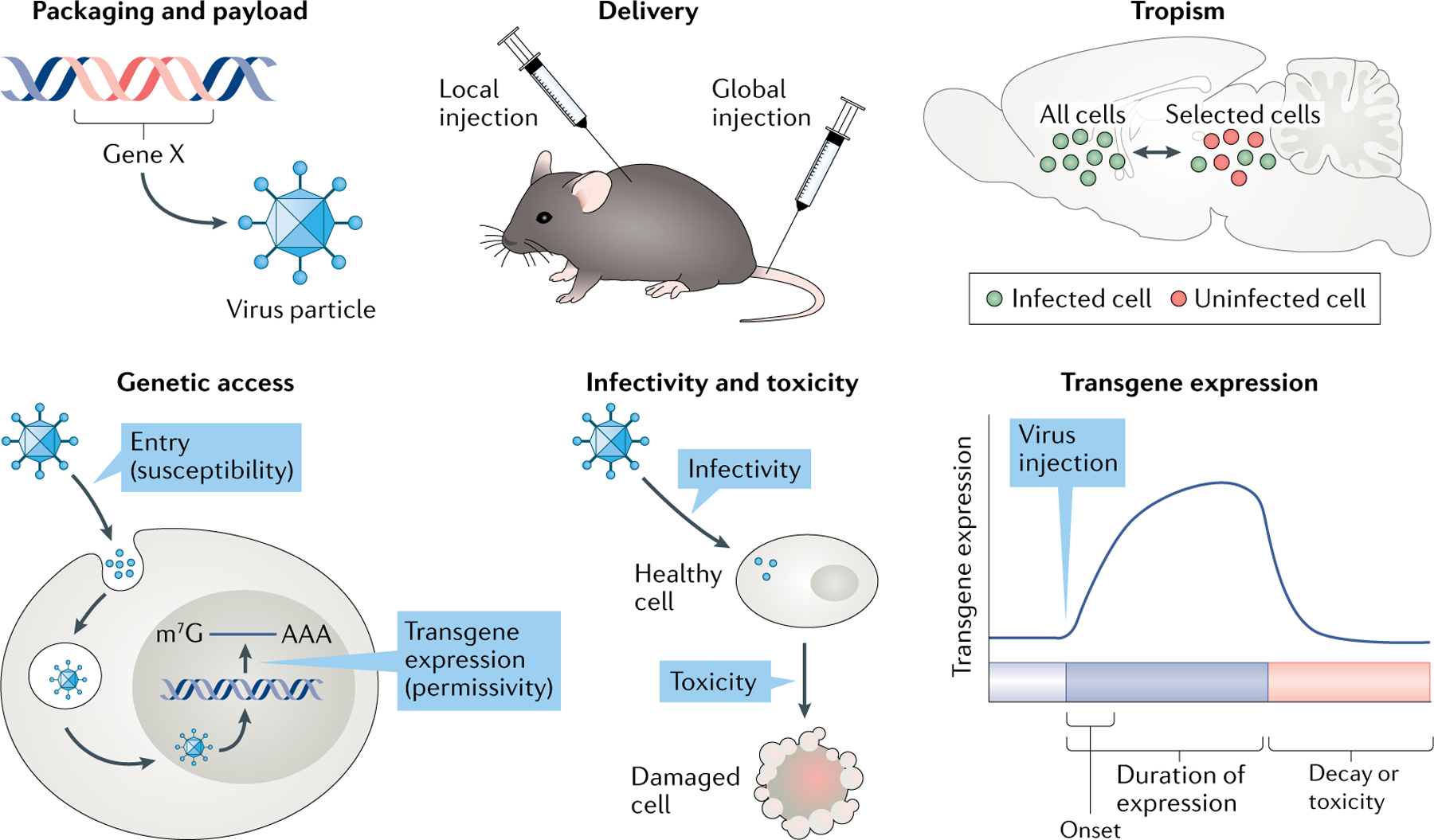
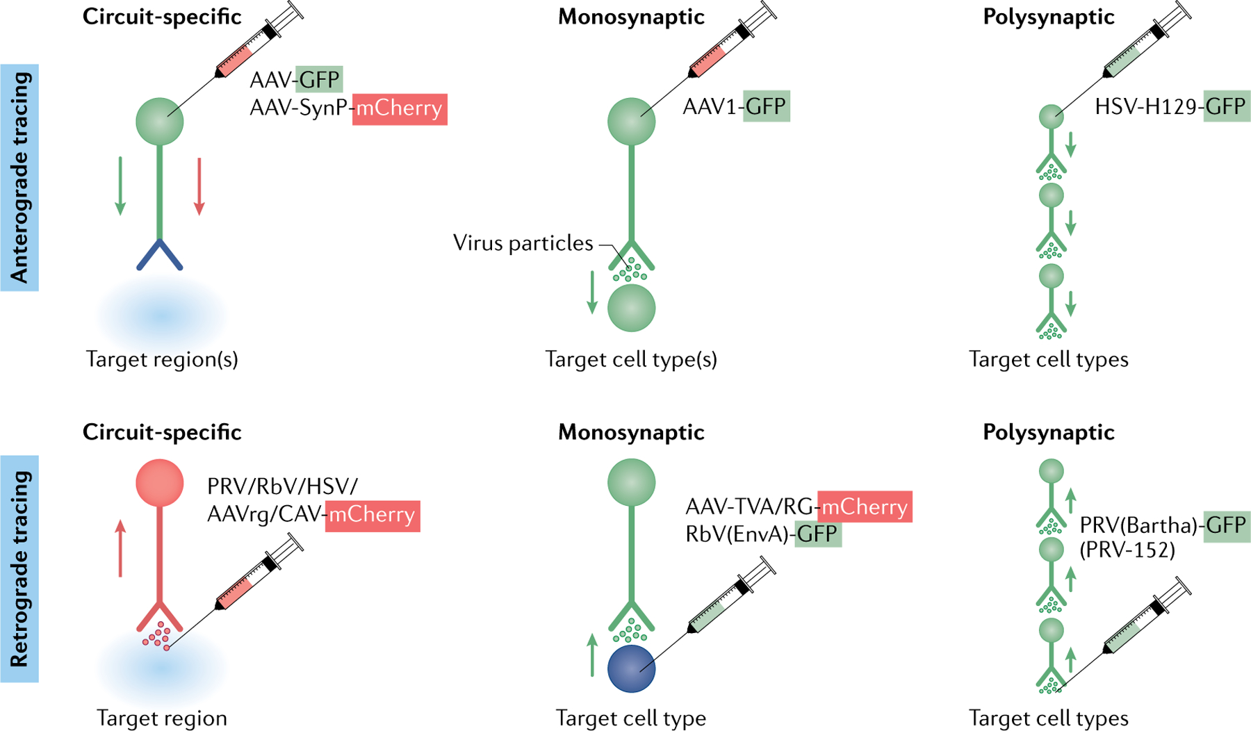
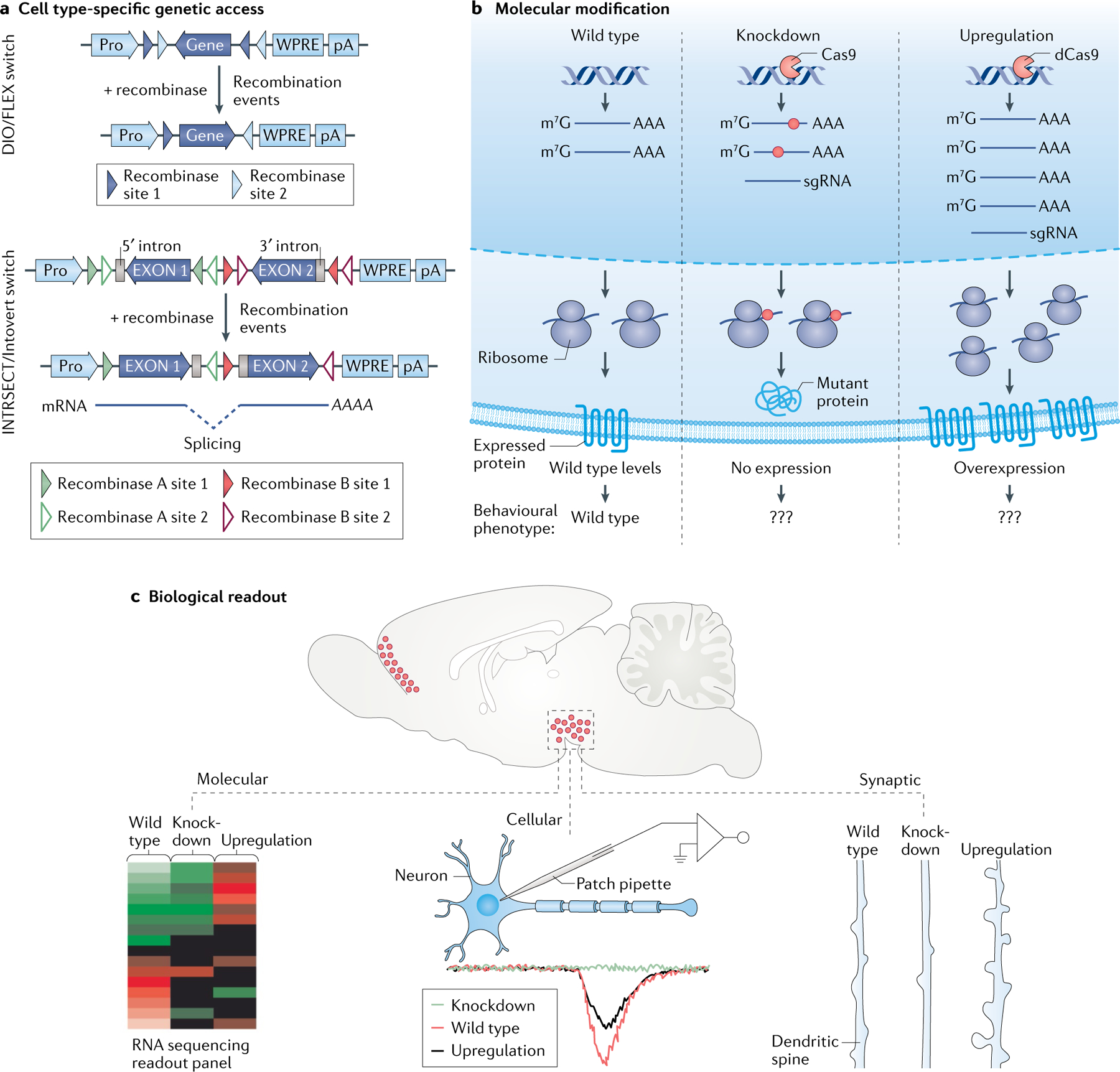
Recombinant viruses are the workhorse of modern neuroscience. Whether one would like to understand a neuron's morphology, natural activity patterns, molecular composition, connectivity or behavioural and physiologic function, most studies begin with the injection of an engineered virus, often an adeno-associated virus or herpes simplex virus, among many other types. Recombinant viruses currently enable some combination of cell type-specific, circuit-selective, activity-dependent and spatiotemporally resolved transgene expression. Viruses are now used routinely to study the molecular and cellular functions of a gene within an identified cell type in the brain, and enable the application of optogenetics, chemogenetics, calcium imaging and related approaches. These advantageous properties of engineered viruses thus enable characterization of neuronal function at unprecedented resolution. However, each virus has specific advantages and disadvantages, which makes viral tool selection paramount for properly designing and executing experiments within the central nervous system. In the current Review, we discuss the key principles and uses of engineered viruses and highlight innovations that are needed moving forward.


Conflict of interest statement

Competing interests

The authors declare no competing interests.

Figures

Fig. 1 |
Fig. 1 |. Key principles for viral-mediated gene transfer in neuroscience.
Schematic demonstrating six key principles essential for the neuroscientist: viral packaging limit (how much nucleic acid a virus particle can carry) and payload (the length and type of genomic material that can be successfully packaged into a virus particle), delivery methods (local versus global injections), tropism (specificity of a virus for a given cell type(s)), access (ability of a virus to enter a cell type and express its gene product(s)), infectivity and toxicity (how efficiently a virus infects a cell and how harmful it is to the cell), and transgene expression dynamics (time course of onset and persistence of transgene expression). These principles play a key role in determining a neuroscientist’s choice of virus by weighing the advantages and disadvantages of a given virus. AAA, 3′ poly(A) tail for mRNA; Gene X, a transgene being packaged into a virus particle; m7G, 7-methylguanosine (5′ cap for mRNA).
Fig. 2 |
Fig. 2 |. Viral strategies for accessing neurons by virtue of their connectivity.
Viral tracing strategies commonly used to gain circuit-specific genetic access to cell types throughout the central nervous system. Tracing methods are commonly divided into anterograde (top) and retrograde (bottom) approaches, which often start by the injection of virus (indicated by a syringe) at a presynaptic soma or at the terminal or postsynaptic soma, respectively. Circuit-specific viral tracing methods most often utilize ‘gutted’ constructs (that is, they express few or no viral genes), which enable access to the cell type with limited toxicity. Monosynaptic and polysynaptic methods for accessing cell types are often, but not always, toxic to infected cells due to the frequent use of live, replication-competent viruses, which are capable of jumping one (monosynaptic) or more (polysynaptic) synapses. Green cell compartments are positive for GFP (green fluorescent protein), and red cell compartments are positive for mCherry (a monomeric, red fluorescent protein). Blue cell compartments (nerve terminals for circuit-specific anterograde tracing and postsynaptic soma in monosynaptic retrograde tracing) are positive for both GFP and mCherry. For circuit-specific anterograde tracing, AAV-GFP and AAV-SynP-mCherry, respectively, help to label the whole cell and the nerve terminals. Note that there is currently no way to access cells that are a given number of synapses upstream or downstream of a target cell type. AAV, adeno-associated virus (AAV1 is a variant of AAV); AAVrg, retrograde-tracing variant of AAV; CAV, canine adenovirus; EnvA, avian ASLV type A envelope protein (cognate ligand for TVA); HSV, herpes simplex virus; HSV-H129, anterograde-tracing variant of HSV; PRV, pseudorabies virus; RbV, rabies virus; RG, rabies G protein; SynP, a synaptophysin fusion construct (can target fluorophores to the presynaptic nerve terminal); TVA, avian receptor for EnvA.
Fig. 3 |
Fig. 3 |. Assessing a gene’s function within the CNS.
Theoretical framework for characterizing the role of a given gene in regulating molecular, cellular, synaptic, circuit and behavioural functions. a | To gain genetic access to a cell population in a cell type-specific fashion, single-recombinase (top) or dual-recombinase (bottom) ‘molecular logic’ can be used. The double-floxed inverted open reading frame (DIO)/flip-excision (FLEX; an irreversible, Cre-dependent molecular switch), often placed before a constitutive promoter (pro), allows for irreversible activation/expression of a cassette. When paired with cell type-specific expression of a recombinase (that is, Cre, Flp, Dre, VCre and so on) using a driver line, the DIO/FLEX switch is irreversibly activated, which leads to constitutive expression of the engineered transgene. This system thus benefits from the specificity of recombinase driver lines and the robust expression of constitutively active promoters (top). INTRSECT (a variant of the FLEX switch; INTRSECT constructs enable intersectional molecular logic through strategic use of introns) and Introvert (a variant of the FLEX switch, using a similar strategy to that of INTRSECT by strategically using introns)-based approaches can also be used to access a cell type using dual-recombinase logic (that is, based on the expression of two recombinases). These recombinases can be used to define any number of features of a cell population, allowing for increased specificity when genetically accessing a cell type (recombinases can be used to access a cell type based on marker gene expression, projection target or activity pattern). Introns are used in the INTRSECT/Introvert approaches to enable strategic inversions of split gene segments (so that the recombinase sites can be spliced out after recombination). This allows for the expansion of new, orthogonal molecular logic operations. Recombinase sites depicted are capable of recombining with one another only if they are of the same colour and shading (bottom). b | Molecular schemata for manipulations of gene expression. Among many options, a gene can be knocked down at the levels of DNA (using, for example, clustered regularly interspaced short palindromic repeats (CRISPR)) or RNA (microRNAs, newer RNA-targeting CRISPR systems). In the example shown, Cas9-mediated CRISPR knockdown is achieved via induction of an insertion-deletion in the genome. This genetic lesion is consequently manifested at the RNA level (represented in the middle sub-panel by a red dot), ultimately resulting in the formation of mutant (non-functional/inactive) protein. For upregulation of endogenous gene expression, a ‘dead’ Cas9 (dCas9) fusion construct enables increased transcription and, ultimately, more protein (represented in the right sub-panel). Triple question marks (???) represent the behavioural phenotype outcome being measured after genetic manipulations. c | Functional readouts after manipulating gene expression. Examples include gene expression adaptations revealed by RNA sequencing. This approach provides a readout of relative expression levels of wild-type, knockdown and upregulated genes (left). Alterations in synaptic strength can be determined using electrophysiology. The sample trace shows hypothetical responses from a neuron with a knockdown, wild-type or overexpressed gene product (middle). At the synaptic level, changes in dendritic spine number and morphology can be compared. These micro-scale adaptations may ultimately lead to observable changes in the animal’s circuit function and behaviour (right). AAA, 3′ poly(A) tail for mRNA; m7G, 7-methylguanosine (5′ cap for mRNA); sgRNA, single guide RNA (enables programmatic targeting of Cas proteins to a desired nucleic acid sequence); WPRE, woodchuck hepatitis virus post-transcriptional regulatory element.

Similar articles

Cited by

References

    1. Baltimore D Expression of animal virus genomes. Bacteriol. Rev 35, 235–241 (1971). - PMC - PubMed
    1. Lefkowitz EJ et al. Virus taxonomy: the database of the International Committee on Taxonomy of Viruses (ICTV). Nucleic Acids Res 46, D708–D717 (2018). - PMC - PubMed
    1. Flint SJ, Racaniello VR, Rall GG, Skalka AM & Enquist LW Principles of Virology 4th edn Vol. 4 (ASM, 2015).
    1. Seo JW et al. Positron emission tomography imaging of novel AAV capsids maps rapid brain accumulation. Nat. Commun 11, 2102 (2020). - PMC - PubMed
    1. Knowland D et al. Distinct ventral pallidal neural populations mediate separate symptoms of depression. Cell 170, 284–297.e18 (2017). - PMC - PubMed