

Modified Le Fort I Osteotomy and Genioplasty for Management of Severe Dentofacial Deformity in β-Thalassaemia Major: Case report and review of the literature
- PMID: 33110654
- PMCID: PMC7574809
- DOI: 10.18295/squmj.2020.20.03.018
Modified Le Fort I Osteotomy and Genioplasty for Management of Severe Dentofacial Deformity in β-Thalassaemia Major: Case report and review of the literature
Abstract
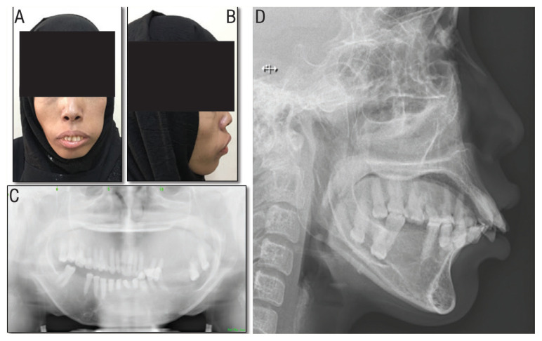
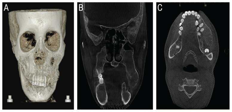
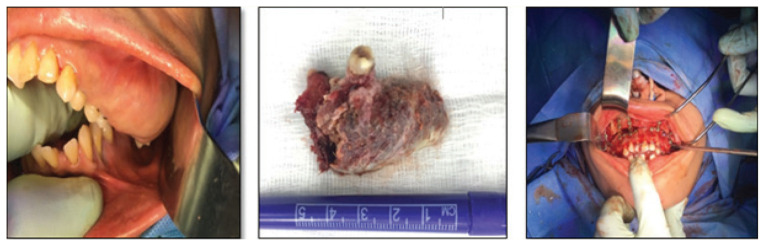
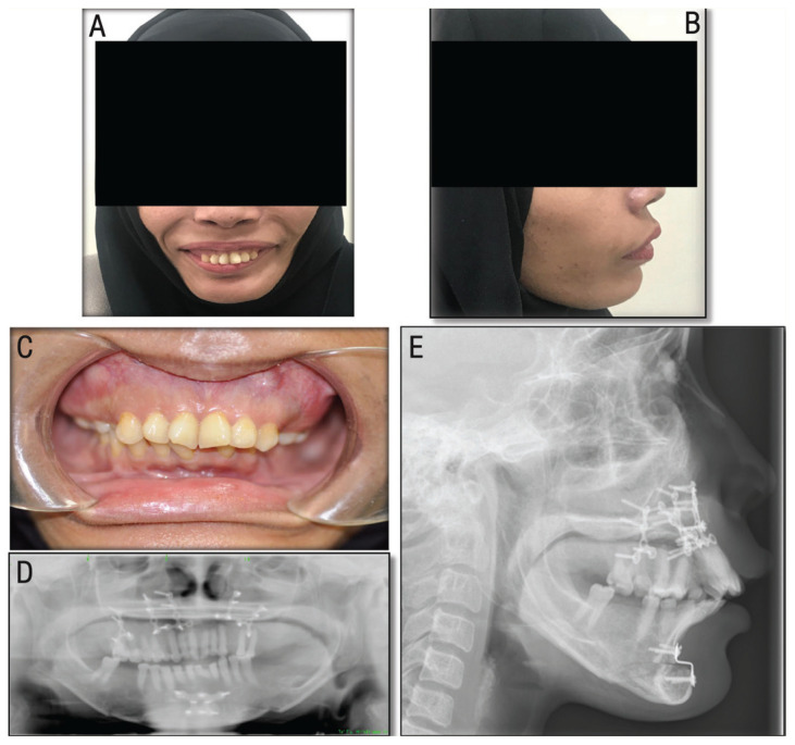
β-thalassaemia major is an autosomal recessive form of haemoglobinopathy that is characterised by complete lack of production of the β-chains resulting in multiple complications that include severe anaemia, failure to thrive and skeletal abnormalities. Facial deformities induced by β-thalassaemia major are rare and are very challenging to treat from a surgical point of view. We report a 33-year-old female patient with β-thalassaemia major who presented to the Dental & Maxillofacial Surgery Department, Sultan Qaboos University Hospital, Muscat, Oman, in 2017 with gross dentofacial skeletal deformity contributing to her psychosocial issues. The facial deformity was corrected surgically by excision of the enlarged maxilla, modified Le Fort I osteotomy and advancement genioplasty. This case highlights the pre-operative preparation, surgical management, encountered complications and treatment outcome within 24 months of follow-up.
Keywords: Beta-Thalassaemia; Case Report; Cooley’s Anemia; Dentofacial Deformities; Genioplasty; Le Fort Osteotomy; Oman; Thalassaemia Major.
© Copyright 2020, Sultan Qaboos University Medical Journal, All Rights Reserved.
Figures
Similar articles
-
Oro-facial characteristics and the surgical correction of patients affected by beta-thalassaemia: a review of the literature and report of a case.Aust Orthod J. 2015 May;31(1):98-106. Aust Orthod J. 2015. PMID: 26219152 Review.
-
Segmental Maxillary Osteotomies in Conjunction With Bimaxillary Orthognathic Surgery: Indications - Safety - Outcome.J Oral Maxillofac Surg. 2016 Jul;74(7):1422-40. doi: 10.1016/j.joms.2016.01.051. Epub 2016 Feb 2. J Oral Maxillofac Surg. 2016. PMID: 26923557
-
Le Fort II Setback Osteotomy to Correct Naso-Ethmoido-Maxillary Protrusion.J Craniofac Surg. 2016 Jan;27(1):e94-9. doi: 10.1097/SCS.0000000000002243. J Craniofac Surg. 2016. PMID: 26674893
-
Management of Maxillary Deformity with Segmental Osteotomy followed by Implant Insertion in β-Thalassemia Major Patient.J Contemp Dent Pract. 2015 Aug 1;16(8):704-7. doi: 10.5005/jp-journals-10024-1744. J Contemp Dent Pract. 2015. PMID: 26423509
-
Blindness as a complication of Le Fort I osteotomy for maxillary distraction.Plast Reconstr Surg. 2002 Feb;109(2):688-98; discussion 699-700. doi: 10.1097/00006534-200202000-00041. Plast Reconstr Surg. 2002. PMID: 11818854 Review.
References
Publication types
MeSH terms
LinkOut - more resources
Full Text Sources