

Sarcoidosis presenting as lacrimal gland enlargement: Eyes speak the truth
- PMID: 33110757
- PMCID: PMC7585470
- DOI: 10.4103/tjo.tjo_125_18
Sarcoidosis presenting as lacrimal gland enlargement: Eyes speak the truth
Abstract
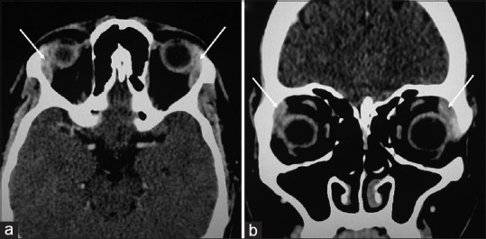
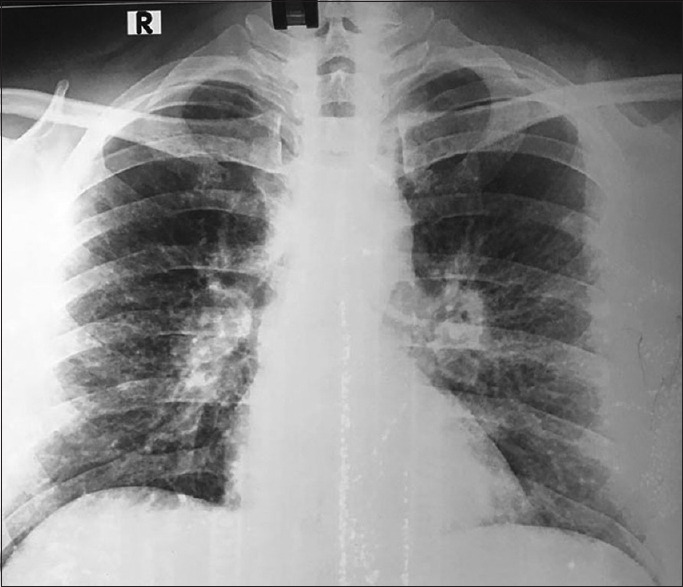
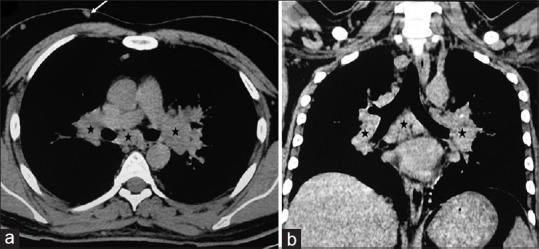
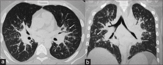
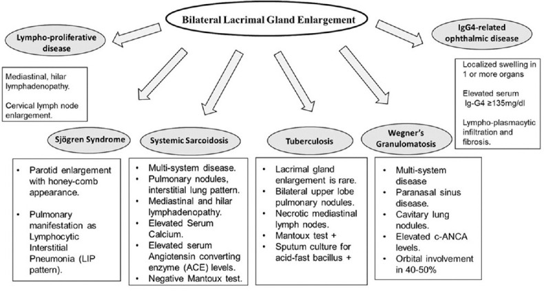
Bilateral lacrimal gland enlargement is uncommon; however, its presence induces brainstorming process and intensive discussion between a clinician and an imaging specialist, leading to exploration of multiple systemic disease patterns such as lymphoproliferative disorders, sarcoidosis, Sjögren's syndrome, and tuberculosis. Thoughtful analysis and diagnostic work-up are required to confirm the diagnosis. Sarcoidosis is a rare systemic disease, with ocular involvement being still rarer. Here, we report a case of a young male presenting with nodular swelling over lateral aspects of both the eyes. The imaging study revealed bilateral lacrimal gland enlargement. Further work-up revealed mediastinal and hilar lymphadenopathy with pulmonary nodules which along with biochemical tests lead to the diagnosis of sarcoidosis. The case highlights the ocular symptoms in sarcoidosis and clinicoradiological approach to bilateral lacrimal gland enlargement.
Keywords: Inflammation; lacrimal gland; sarcoidosis.
Copyright: © 2019 Taiwan J Ophthalmol.
Conflict of interest statement
The authors declare that there are no conflicts of interest in this paper.
Figures
References
-
- American Academy of Ophthalmology. Sarcoidosis. [[Last accessed on 2019 Jan 15]]. Available from: https://wwwaaoorg/bcscsnippetdetailaspxid=7666e4b7-664f-477d-af8b-0ef1aa... .
-
- Manavi K, Skouroumouni G, Papaderakis G, Panagiotidou D, Tsitouridis D. Rare Case of Sarcoidosis Involving Primarily the Parotid Glands: US and MRI Correlation. 2017. [[Last accessed on 2018 Nov]]. Available from: http://wwweuroradorg/casephpid=14570 .
-
- Usui Y, Kaiser ED, See RF, Rao NA, Sharma OP. Update of ocular manifestations in sarcoidosis. Sarcoidosis Vasc Diffuse Lung Dis. 2002;19:167–75. - PubMed
-
- Farmer JP, Lamba M, Lamba WR, Jordan DR, Gilberg S, Sengar DP, et al. Lymphoproliferative lesions of the lacrimal gland: Clinicopathological, immunohistochemical and molecular genetic analysis. Can J Ophthalmol. 2005;40:151–60. - PubMed
Publication types
LinkOut - more resources
Full Text Sources