. 2020 Oct 14:2020:8256809.
doi: 10.1155/2020/8256809. eCollection 2020.

In Vitro Anticancer Potential of Berberis lycium Royle Extracts against Human Hepatocarcinoma (HepG2) Cells

Affiliations

In Vitro Anticancer Potential of Berberis lycium Royle Extracts against Human Hepatocarcinoma (HepG2) Cells

Kiren Mustafa et al. Biomed Res Int. .

Abstract

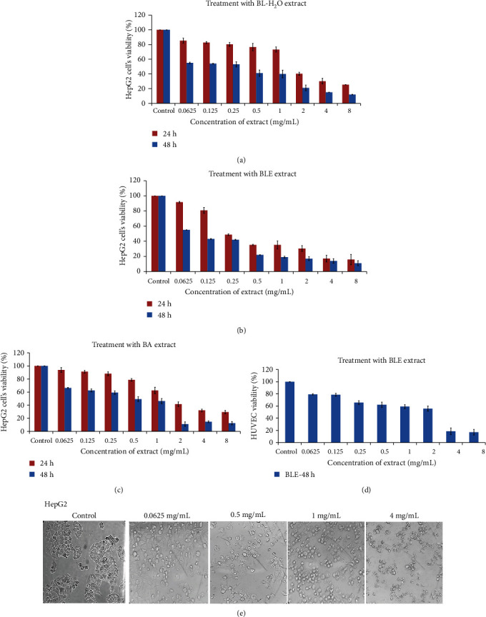
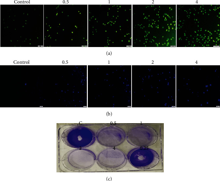
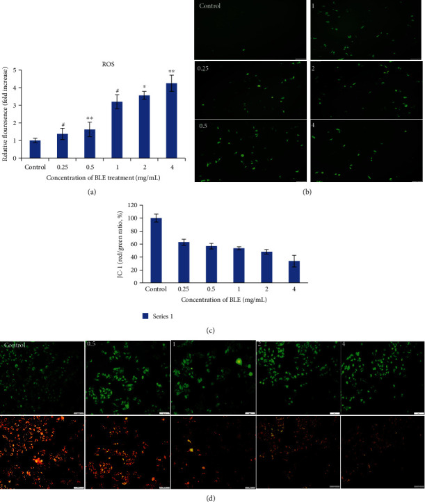
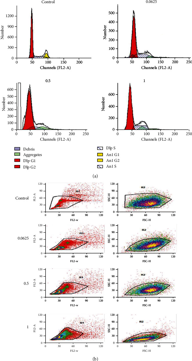
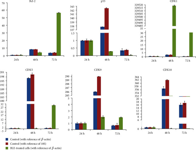
Human liver cancer has emerged as a serious health concern in the world, associated with poorly available therapies. The Berberis genus contains vital medicinal plants with miraculous healing properties and a wide range of bioactivities. In this study, different crude extracts of B. lycium Royle were prepared and screened against Human Hepatocarcinoma (HepG2) cell lines. The water/ethanolic extract of B. lycium Royle (BLE) exhibited significant antiproliferative activity against the HepG2 cancer cell line with an IC50 value of 47 μg/mL. The extract decreased the clonogenic potential of HepG2 cells in a dose-dependent manner. It induced apoptotic cell death in HepG2 cells that were confirmed by cytometric analysis and microscopic examination of cellular morphology through DAPI-stained cells. Biochemical evidence of apoptosis came from elevating the intracellular ROS level that was accompanied by the loss of mitochondrial membrane potential. The mechanism of apoptosis was further confirmed by gene expression analysis using RT-qPCR that revealed the decline in Bcl-2 independent of p53 mRNA and a rise in CDK1 while downregulating CDK5, CDK9, and CDK10 mRNA levels at 48 h of BLE treatment. The most active fraction was subjected to HPLC which indicated the presence of berberine (48 μg/mL) and benzoic acid (15.8 μg/mL) as major compounds in BLE and a trace amount of luteolin, rutin, and gallic acid. Our study highlighted the importance of the most active BLE extract as an excellent source of nutraceuticals against Human Hepatocarcinoma that can serve as an herbal natural cure against liver cancer.


Conflict of interest statement

The authors declare that there is no conflict of interest regarding the publication of this paper.

Figures

Figure 1
Figure 1
Time- and dose-dependent inhibitory effect of B. Lycium Royle extracts on HepG2 cells and HUVECs. (a) Water extract on the cell's viability for 24 and 48 hours. (b) Effect of 50% ethanol extract of BLE on HepG2 cell's growth for 24 and 48 h. (c) BL acetone extract decreased the viability of HepG2 cells in a dose- and time-dependent manner. (d) Inhibitory effect of BL water extract on HUVECs as observed for a maximum of 48 h. (e) Morphological examination of HepG2 cells after 24 h of treatment with BL-50% ethanol extract, with a light microscope (only a few photographs have been shown here with a significant difference). All values are presented as mean ± SD from three independent experiments.
Figure 2
Figure 2
Fluorescent microscopy of HepG2 cells and colony formation assay. (a) AO/EB staining of HepG2 cells after incubation with 0.5, 1, 2, and 4 mg/mL of BLE for 24 h. Live cells stained as uniformly light green in control (untreated) cells, while early apoptotic cells stained as green cells with bright green nuclei that gives the bright green appearance, and necrotic cells/late apoptotic cells stained as light orange. (b) DAPI staining shows the control (untreated) cells as uniformly light blue and the cells after 24 hour of incubation with BLE appeared bright blue with dense nuclei due to chromosomal condensation and nuclear fragmentation as a result of apoptosis. All pictures were taken under the fluorescence microscope (scale bar = 20 μm). (c) The HepG2 cells were grown on a 6-well plate and subsequently incubated with BLE for 24 h, followed by 8-day growth in complete media; it exhibited complete disassociation of colony-forming potential of cancer cells at all high (4, 2, 1, and 0.5 mg/mL) doses and its ability diminishes at 0.25 mg/mL.
Figure 3
Figure 3
Effect of BLE on ROS generation and mitochondrial membrane potential changes in HepG2 cells. HepG2 cells were incubated without or with BLE at specified concentrations for 24 hours. (a) The generation of ROS was analyzed by DCFH-DA (10 μM/L) assay in HepG2 cells, and the fluorescence was measured at excitation and emission λ of 488 and 525 nm, respectively, with the help of a fluorescence microplate reader. (b) Fluorescence microscopic view of HepG2 cells after ROS assay indicates an increase in green fluorescence spots due to ROS generation with BLE treatment as compared to control. (c) The bar graph represents the fluorescent intensity ratios between green fluorescence (for loss of membrane potential at 485 nm-535 nm λ) and red fluorescence (for normal changes in mitochondrial membrane potential at 535 nm-590 nm λ) by the fluorescent microplate reader. The JC-1 red fluorescence intensity was selected as 100%, and fluorescence intensity of treated samples was measured relative to control as (treated/control) × 100. (d) Florescent microscopic view of HepG2 cells for Δψ after staining in the dark with a JC-1 probe for 30 min and red fluorescence was present in cells with high ψ and vice versa for green fluorescence; photographs were taken at two λ (green and red). All values are mean ± SD from three independent experiments. Significant differences as compared to control were depicted by p, 0.05; ∗∗p, 0.01; or ∗∗∗p, 0.001.
Figure 4
Figure 4
Selective arrest of the cell cycle by BLE against HepG2 cells after 24 h of incubation. (a) The histograms were made by analysis on a ModFit LT model of analysis for control and BLE-treated HepG2 cells at mentioned concentrations for 24 h. BLE arrested the HepG2 cell's cycle at the synthesis (S) phase of growth at 0.0625 mg/mL and G1 phase after treatment at high concentrations of 0.5 and 1 mg/mL. (b) Gate 1 is plotted with FL2-W (width) and FL2-A (area) of the scattered cells, and gate 2 defines FSC-H (forward scatter) and SSC-H (side scatter) of the stained HepG2 cells.
Figure 5
Figure 5
Variable genetic expression of key apoptotic/cell cycle markers after treatment with BLE. The mRNA expression is relative to 24 h. All results are stated as mean ± SD from experiments repeated three times.

References

    1. El-Serag H. B. Hepatocellular carcinoma. The New England Journal of Medicine. 2011;365(12):1118–1127. doi: 10.1056/NEJMra1001683. - DOI - PubMed
    1. Yang J. D., Roberts L. R. Hepatocellular carcinoma: a global view. Nature Reviews Gastroenterology & Hepatology. 2010;7(8):448–458. doi: 10.1038/nrgastro.2010.100. - DOI - PMC - PubMed
    1. Spangenberg H. C., Thimme R., Blum H. E. Targeted therapy for hepatocellular carcinoma. Nature Reviews Gastroenterology & Hepatology. 2009;6(7):423–432. doi: 10.1038/nrgastro.2009.86. - DOI - PubMed
    1. Mokhber-Dezfuli N., Saeidnia S., Gohari A. R., Kurepaz-Mahmoodabadi M. Phytochemistry and pharmacology of berberis species. Pharmacognosy Reviews. 2014;8(15):8–15. doi: 10.4103/0973-7847.125517. - DOI - PMC - PubMed
    1. Arayne M. S., Sultana N., Bahadur S. S. The Berberis story: Berberis vulgaris in therapeutics. Pakistan Journal of Pharmaceutical Sciences. 2007;20(1):83–92. - PubMed

MeSH terms