

doi: 10.1016/j.xpro.2020.100086.
eCollection 2020 Sep 18.
Protocol for Primary Mouse Hepatocyte Isolation
Affiliations
- PMID: 33111119
- PMCID: PMC7580103
- DOI: 10.1016/j.xpro.2020.100086
Item in Clipboard
Protocol for Primary Mouse Hepatocyte Isolation
STAR Protoc.
.
Abstract
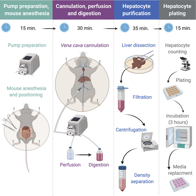
Primary hepatocytes are a vital tool in various biomedical research disciplines, serving as an ex vivo model for liver physiology. Obtaining high yields of viable primary mouse hepatocytes is technically challenging, limiting their use. Here, we present an improved protocol based on the classic two-step collagenase perfusion technique. The liver is washed by perfusion, hepatocytes are dissociated by collagenase, separated from other cells, and cultured. This protocol was optimized to significantly reduce procedure duration and improve hepatocyte yield and viability.
© 2020 The Author(s).
Conflict of interest statement
The authors declare no competing interests.
Figures
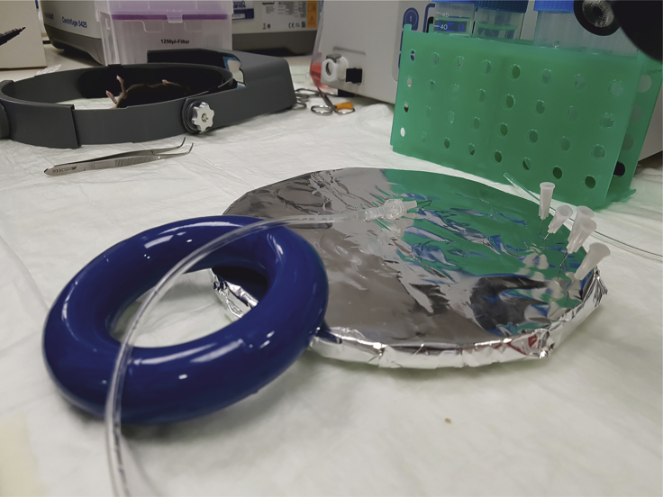
Needle and Tubing Preparation The needle is connected to the outlet end of the tubing by using a luer lock. The tubing and needle fixture is supported by a heavy object (here a media bottle ring weight) to elevate it from the dissection tray. This elevation facilitates proper cannulation.
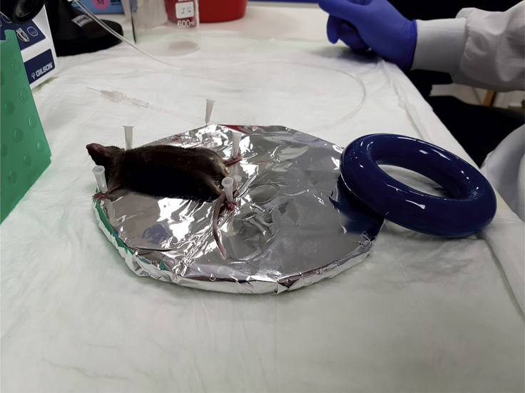
Mouse Positioning on the Dissection Tray The anaesthetized mouse is placed on the edge of the dissection tray with its head protruding outside and its limbs secured with needles. Securing limbs with tape is not recommended because the area will be soaked with liquid during procedure, potentially loosening adhesion.
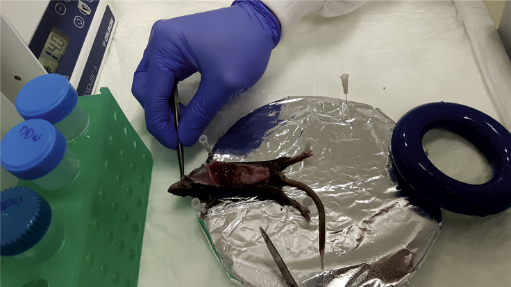
Incision and Preparing for Cannulation The mouse fur and skin are cut in a “U” shape, the mouse skin is placed and secured near the head with a needle.
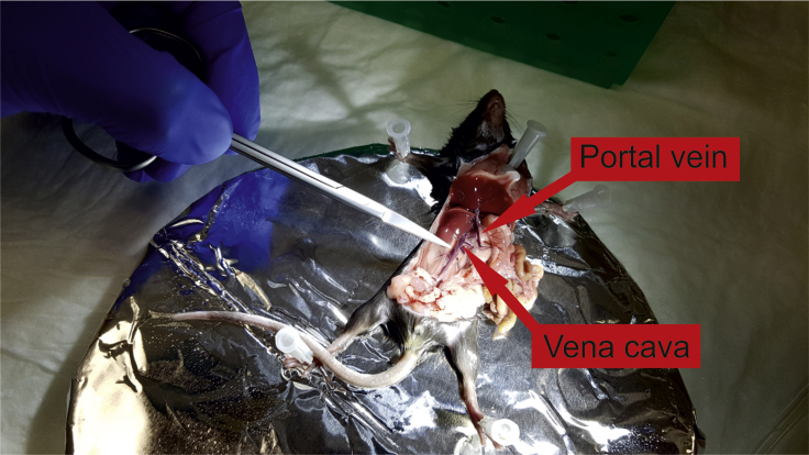
Portal Vein and Vena Cava exposure Mouse intestine and the rest of the viscera are moved to the right. Both the portal vein and vena cava are revealed.
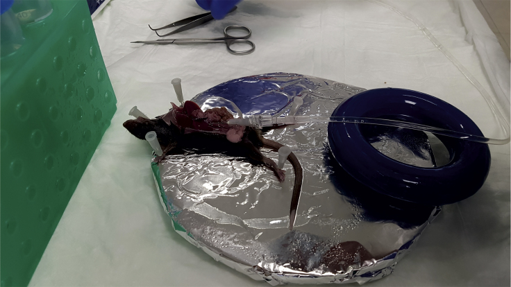
Cannulation of the Vena Cava The tip of the needle is inserted (bevel side up, in an almost flat angle) into the vena cava above the kidney. The tubing rests on the elevating object without manual support.
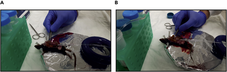
Clamping the Portal Vein (A and B) The portal vein is clamped with forceps for 7–10 s to stop fluid exiting the liver. Liver swells upon clamping (compare A to B).
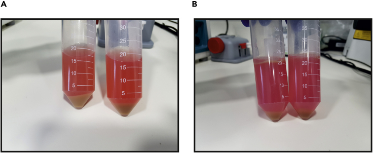
Hepatocyte Purification (A) Cells from the dissected liver are released into suspension and centrifuged. The pellet contains hepatocytes and the rest of the cells are left in the supernatant. (B) The hepatocyte pellet (A) is re-suspended with Percoll solution and centrifuged again. The pellet contains viable hepatocytes while dead cells and debris are left in the supernatant.
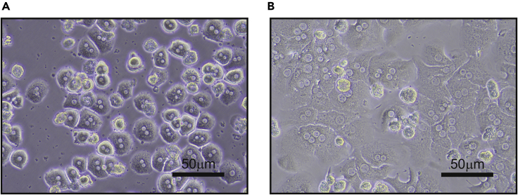
Hepatocyte Plating (A) Hepatocytes are plated and after 3 h have adhered to the surface, acquiring a spherical shape. (B) Twenty-four hours following plating hepatocytes acquire their typical hexagonal shape.
References
-
- Azimifar S.B., Nagaraj N., Cox J., Mann M. Cell-type-resolved quantitative proteomics of murine liver. Cell Metab. 2014;20:1076–1087. - PubMed
-
- Casciano D.A. Development and utilization of primary hepatocyte culture systems to evaluate metabolism, DNA binding, and DNA repair of xenobiotics. Drug Metab. Rev. 2000;32:1–13. - PubMed
Publication types
MeSH terms
Substances
Grants and funding
LinkOut - more resources
Full Text Sources
