. 2020 Nov 24;20(23):4486-4501.
doi: 10.1039/d0lc00875c.

Modeling ascending infection with a feto-maternal interface organ-on-chip

Affiliations

Modeling ascending infection with a feto-maternal interface organ-on-chip

Lauren S Richardson et al. Lab Chip. .

Abstract

Maternal infection (i.e., ascending infection) and the resulting host inflammatory response are risk factors associated with spontaneous preterm birth (PTB), a major pregnancy complication. However, the path of infection and its propagation from the maternal side to the fetal side have been difficult to study due to the lack of appropriate in vitro models and limitations of animal models. A better understanding of the propagation kinetics of infectious agents and development of the host inflammatory response at the feto-maternal (amniochorion-decidua, respectively) interface (FMi) is critical in curtailing host inflammatory responses that can lead to PTB. To model ascending infection and determine inflammatory responses at the FMi, we developed a microfluidic organ-on-chip (OOC) device containing primary cells from the FMi (decidua, chorion, and amnion [mesenchyme and epithelium]) and collagen matrix harvested from primary tissue. The FMi-OOC is composed of four concentric circular cell/collagen chambers designed to mimic the thickness and cell density of the FMi in vivo. Each layer is connected by arrays of microchannels filled with type IV collagen to recreate the basement membrane of the amniochorion. Cellular characteristics (viability, morphology, production of nascent collagen, cellular transitions, and migration) in the OOC were similar to those seen in utero, validating the physiological relevance and utility of the developed FMi-OOC. The ascending infection model of the FMi-OOC, triggered by exposing the maternal (decidua) side of the OOC to lipopolysaccharide (LPS, 100 ng mL-1), shows that LPS propagated through the chorion, amnion mesenchyme, and reached the fetal amnion within 72 h. LPS induced time-dependent and cell-type-specific pro-inflammatory cytokine production (24 h decidua: IL-6, 48 h chorion: GM-CSF and IL-6, and 72 h amnion mesenchyme and epithelium: GM-CSF and IL-6). Collectively, this OOC model and study successfully modeled ascending infection, its propagation, and distinct inflammatory response at the FMi indicative of pathologic pathways of PTB. This OOC model provides a novel platform to study physiological and pathological cell status at the FMi, and is expected to have broad utility in the field of obstetrics.


Conflict of interest statement

Conflict of interest statement: A patent application describing the feto-maternal interface organ-on-a-chip system has been submitted.

Figures

Figure 1:
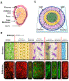
Figure 1:. Recreating the FMi with organ-on-chip technology.
A) An illustration of the anatomy of the intrauterine tissue showing maternal and fetal components and both FMis (black arrows [black box: placenta-decidua basalis; white box: fetal membrane – decidua parientalis]). Maternal tissues are comprised of the uterus (myometrium) and cervix, while the fetal tissues include the placenta, umbilical cord, fetus, and fetal membrane (amniochorionic membrane). B) Region of the decidua and amniochorionic membrane indicated as a white box in A, showing the FMi of interest. The FMi is divided by maternal decidual cells (black text) and fetal-derived amniochorionic cells (gray text). The top row shows a schematic of the amniochorionic membrane and maternal decidua. From left to right, the description starts from the outermost layer (the maternal decidua) and ends at the fetal amnion. The chorion interfaces with the maternal decidua (green), thus connecting the fetal layers to the maternal compartment of the uterus. The chorion (yellow) is connected to the ECM through a type IV collagen basement membrane (green stripes) and is made up of chorion trophoblasts (CTs). The fibroblast, spongy, and reticular layers of the ECM harbor amnion mesenchymal cells (AMCs) and chorion mesenchymal cells (CMCs) (purple). The AECs (blue) are connected to the ECM through the basement membrane/compact layer (green stripes). The bottom row shows corresponding multiphoton microscopy images of each section, where cells are stained in red color and the collagen layers are stained in green color. The red lines divide the four different compartments of the FMi that are represented in the FMi-OOC. Scale bar = 30 μm. C) Schematic of the FMi-OOC device designed to mimic the FMi. The FMi-OOC is composed of four concentric circular cell culture chambers separated by arrays of microchannels. The cells are seeded as follows, from the center to the outside: decidua cells (green), CMCs/CTs (yellow), AMCs – amnion mesenchyme (purple), and AECs – amnion epithelium (blue), respectively. Primary fetal membrane collagen and Matrigel (pink) can enable culturing AMCs and CMC/CTs in a 3D format. To recreate cell-collagen interfaces, the microchannels are filled with type IV collagen (pink) to mimic the basement membrane of the amnion and chorion layers. In contrast, the choriodecidua interface was not filled with collagen (gray).
Figure 2:
Figure 2:. Design and images of the FMi-OOC model.
A) Schematic illustration of the FMi-OOC highlighting the four cell culture chambers connected by arrays of 24 microchannels. B) Cross-sectional view of the cell culture chambers showing the chamber height (250 μm), individual chamber diameters (mimicking in utero thickness), and the interconnecting microchannel lengths. C) Image of the microfabricated FMi-OOC where each cell culture chamber were filled with different color dye for easy visualization, namely the choriodecidua interface (decidua – green; chorion – yellow) and the amniochorionic interface (chorion – yellow; amnion – AMC [purple] and AEC [blue]). D) Design of an on-chip media reservoir layer that was aligned and placed on top of the cell loading inlets and outlets (seen in image C) of the main cell culture layer, both to provide sufficient media for culture and also to control the diffusion between the cell culture layer through controlling the hydrostatic pressure between the chambers by filling the reservoirs to different height. I addition, localized chemical treatments can be performed as well as effluents collected from each culture chamber through these reservoirs. The center decidua layer has one reservoir, the chorion and AMC have two each, while the AEC chamber has four. E) The FMi-OOC device with the integrated media reservoir filled with color dye in each of the corresponding cell culture layers.
Figure 3:
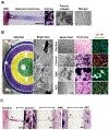
Figure 3:. Characterization of decidua and amniochorionic derived cells cultured within the FMi-OOC.
A) Microchannels between the AEC and AMC, as well as the AMC and CTs/CMCs, chambers are filled with type IV collagen to recreate the two basement membranes. Collagen was stained with Masson trichome for visualization (blue color, left image). Scale bar = 100 μm. Additionally, AMCs and CTs/CMCs were cultured with decellularized primary collagen (middle image) harvested from the membrane of the amnion to provide biochemical factors necessary to recreate stromal layers, as well as function as and matrix to provide a scaffold for the cells to grow in 3D (right image). B) On the left is a cross-sectional view of the four-chamber FMi-OOC device, highlighting the decidua chamber as green, the chorion chamber as yellow, the AMC chamber as purple, and the AEC chamber as blue. The right section of this image shows bright field microscopy images of cells growing in each cell culture chamber. A variety of in utero characteristics were measured to determine if cells grown within the FMi-OOC retained their in vivo characteristics (n = 3). These measurements included cell morphology, collagen production (Masson trichome staining [non-collagen producing cells show up as red, collagen-producing cells show up as purple, and collagen components show up as dark purple/blue]), cellular transition status (epithelial [CK-18; red] and mesenchymal [vimentin; green]) intermediate filament expression, and migratory potential. Scale bar = 50 μm. C) Visualization of cell migration between compartments through the use of Masson trichome stain as a counter stain. Cellular migration between the chorion and amnion chambers were observed (see black arrowhead), as well as degradation of type IV collagen in the microchannel within 72 h. Scale bar = 100 μm.
Figure 4:
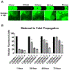
Figure 4:. Fluidic isolation and diffusion of florescent LPS over time between the FMi-OOC chambers.
A) FITC-LPS was tracked every 12 h for 72 h to monitor its diffusion from the maternal decidua side (chamber 1) to the fetal AEC side (chamber 4). Scale bar = 600 μm. B) FITC-LPS significantly propagated between each chamber in the 24 h time increment: chamber 1 (1 h [85±0 RFU] → 24 h [83±0.5 RFU]), chamber 2 (1 h [20±0.6 RFU] → 24 h [53±6 RFU] [P<0.0001]), chamber 3 (24 h [27±3 RFU] → 48 h [50±4 RFU] [P<0.0001]), and chamber 4 (48 h [27±3 RFU] → 72 h [42±3 RFU] [P=0.0074]) (n = 3). Values are expressed as mean intensities ± SEM.
Figure 5:
Figure 5:. Propagation of LPS in the FMi-OOC.
Immunocytochemistry followed by fluorescent microscopy shows LPS (red dots; white arrows) propagation from the decidua (24 h – 0.09±0.02 RFI), to chorion (24 h – 0.005±0.001 RFI), and then to AMCs (48 h – 0.009±0.006 RFI) within 48 h, and finally to AECs within 72 h (72 h – 0.01±0.003 RFI) (n = 3). Scale bar = 10 μm. Blue – DAPI, Red – LPS. Values are expressed as mean intensities ± SEM.
Figure 6:
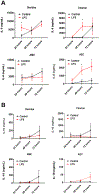
Figure 6:. Production and propagation of pro- and anti-inflammatory cytokines in the FMi-OOC device.
A) Increased IL-6 level in decidual cells after 24-h LPS treatment can be observed compared to controls (P=0.0287). The chorion and AECs also secreted significant levels of IL-6 after 48 h of LPS treatment (P=0.0261 and P=0.0161, respectively). This trend continued to increase in the AEC compartment after 72 h (P=0.0376), showing a progressive induction of inflammatory mediators from the decidua to AEC (n = 4). B) LPS decreased the production of the anti-inflammatory IL-10 in the decidua, chorion, and AMCs at all time points compared to controls. However, LPS significantly increased IL-10 levels in AECs after 72 h (P=0.0376) (n = 4). For all graphs, values are expressed as mean values ± SEM.
Figure 7:
Figure 7:. Schematic showing the propagation of infection and inflammation across the FMi observed through the FMi-OOC model.
Using the developed FMi-OOC model, an ascending infection model (decidua → amnion) of microbial invasion of the amniotic cavity was recreated. Ascending infection was shown associated with a pro-inflammatory shift, compromising the FMi immune homeostasis that can eventually result in intra-amniotic inflammation predisposing subjects to preterm labor-associated feto-maternal inflammatory responses. A) Illustration of ascending infection during pregnancy that the developed model recapitulates. B) The FMi (decidua-fetal amniochorionic interface) interconnects the cervix and intra-amniotic cavity. During ascending infection, infectious microbes transverse through the cervix, propagate from the decidua to reach AECs, and initiate inflammation. C) Propagation of infection coincides with an increased pro-inflammatory environment (high IL-6 and GM-CSF levels; low IL-10 level), causing an immune imbalance.

Similar articles

Cited by

References

    1. Keelan JA, J Reprod Immunol, 2018, 125, 89–99. - PubMed
    1. Romero R, Dey SK and Fisher SJ, Science, 2014, 345, 760–765. - PMC - PubMed
    1. Menon R and Fortunato SJ, Best Pract Res Clin Obstet Gynaecol, 2007, 21, 467–478. - PubMed
    1. Bastek JA, Gomez LM and Elovitz MA, Clin. Perinatol, 2011, 38, 385–406. - PubMed
    1. Behnia F, Sheller S and Menon R, Am. J Reprod Immunol, 2016, DOI: 10.1111/aji.12496 [doi]. - DOI - PubMed

Publication types

Substances