

Nutrient sensing in Leishmania: Flagellum and cytosol
- PMID: 33112443
- PMCID: PMC8087152
- DOI: 10.1111/mmi.14635
Nutrient sensing in Leishmania: Flagellum and cytosol
Abstract
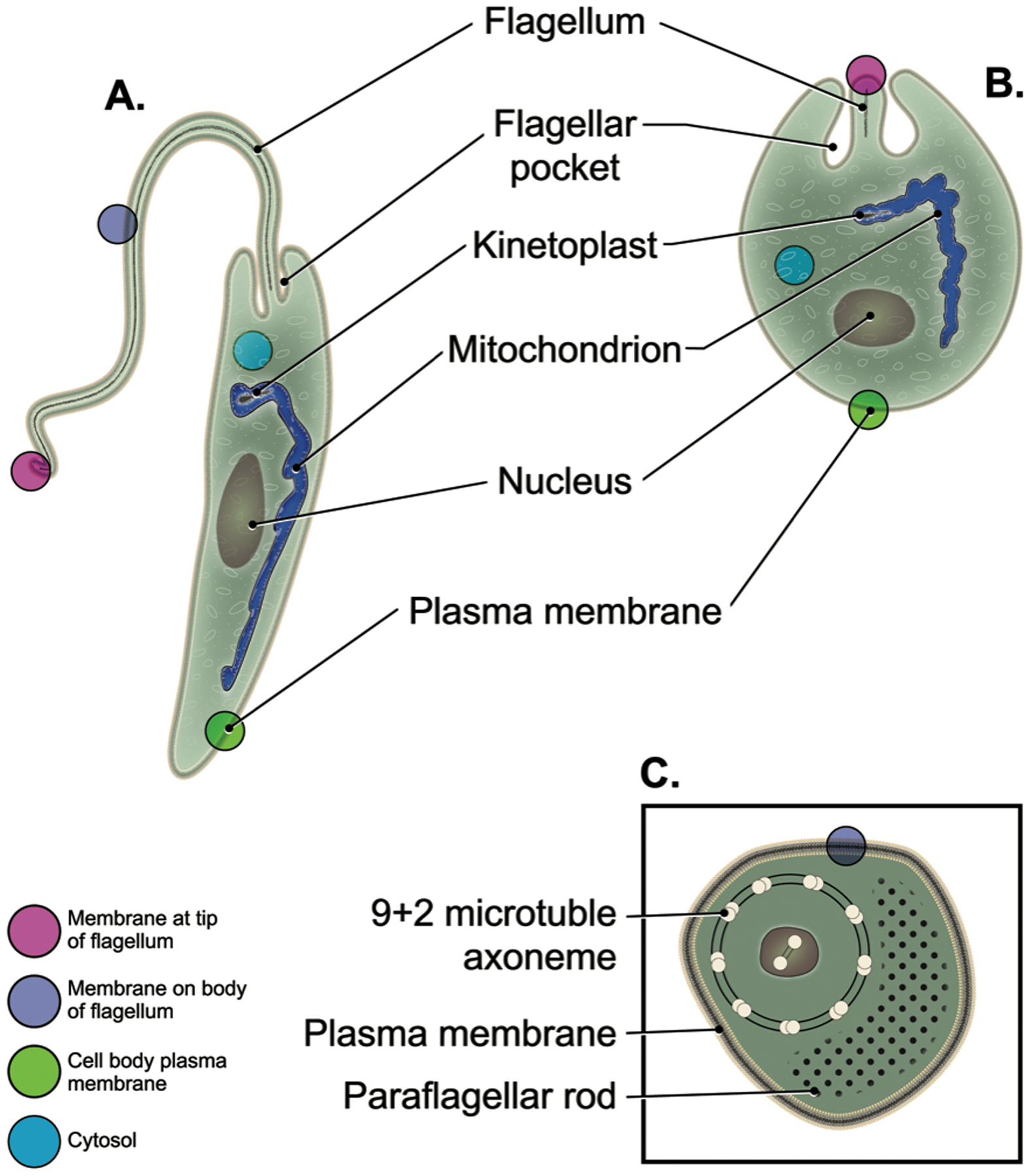
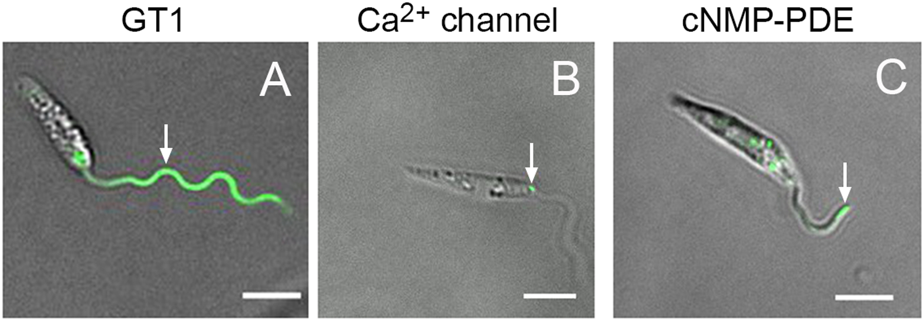
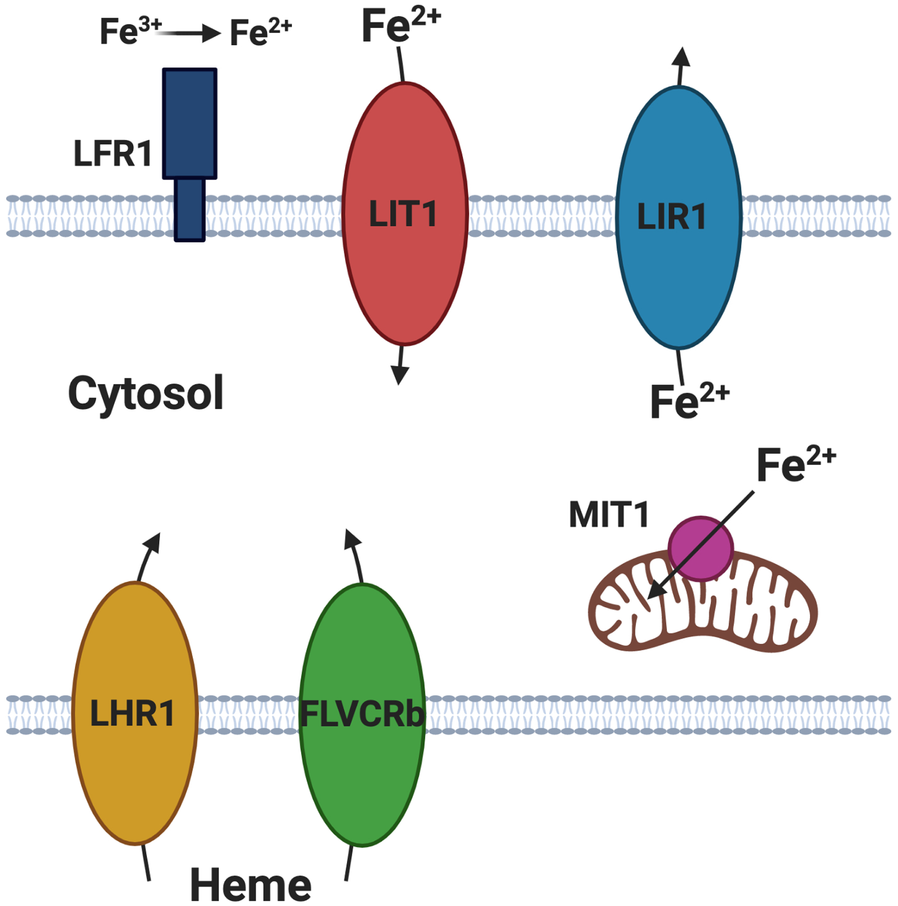
Parasites are by definition organisms that utilize resources from a host to support their existence, thus, promoting their ability to establish long-term infections and disease. Hence, sensing and acquiring nutrients for which the parasite and host compete is central to the parasitic mode of existence. Leishmania are flagellated kinetoplastid parasites that parasitize phagocytic cells, principally macrophages, of vertebrate hosts and the alimentary tract of sand fly vectors. Because nutritional supplies vary over time within both these hosts and are often restricted in availability, these parasites must sense a plethora of nutrients and respond accordingly. The flagellum has been recognized as an "antenna" that plays a core role in sensing environmental conditions, and various flagellar proteins have been implicated in sensing roles. In addition, these parasites exhibit non-flagellar intracellular mechanisms of nutrient sensing, several of which have been explored. Nonetheless, mechanistic details of these sensory pathways are still sparse and represent a challenging frontier for further experimental exploration.
© 2020 John Wiley & Sons Ltd.
Conflict of interest statement
Conflict of interest
The authors declare no financial conflict of interest.
Figures
References
-
- Alcolea PJ, Alonso A, Baugh L, Paisie C, Ramasamy G, Sekar A, Sur A, Jimenez M, Molina R, Larraga V, and Myler PJ (2018) RNA-seq analysis reveals differences in transcript abundance between cultured and sand fly-derived Leishmania infantum promastigotes. Parasitol Int 67: 476–480. - PubMed
-
- Beneke T, Demay F, Hookway E, Ashman N, Jeffery H, Smith J, Valli J, Becvar T, Myskova J, Lestinova T, Shafiq S, Sadlova J, Volf P, Wheeler RJ, and Gluenz E (2019) Genetic dissection of a Leishmania flagellar proteome demonstrates requirement for directional motility in sand fly infections. PLoS Pathog 15: e1007828. - PMC - PubMed
-
- Bhattacharya A, Biswas A, and Das PK (2009) Role of a differentially expressed cAMP phosphodiesterase in regulating the induction of resistance against oxidative damage in Leishmania donovani. Free Radic Biol Med 47: 1494–1506. - PubMed
-
- Cabello-Donayre M, Malagarie-Cazenave S, Campos-Salinas J, Galvez FJ, Rodriguez-Martinez A, Pineda-Molina E, Orrego LM, Martinez-Garcia M, Sanchez-Canete MP, Estevez AM, and Perez-Victoria JM (2016) Trypanosomatid parasites rescue heme from endocytosed hemoglobin through lysosomal HRG transporters. Mol Microbiol 101: 895–908. - PubMed
Publication types
MeSH terms
Substances
Grants and funding
LinkOut - more resources
Full Text Sources
Medical
