. 2021 Apr;19(4):767-784.
doi: 10.1111/pbi.13503. Epub 2020 Nov 28.

Overexpression of alfalfa SIMK promotes root hair growth, nodule clustering and shoot biomass production

Affiliations

Overexpression of alfalfa SIMK promotes root hair growth, nodule clustering and shoot biomass production

Miroslava Hrbáčková et al. Plant Biotechnol J. 2021 Apr.

Abstract

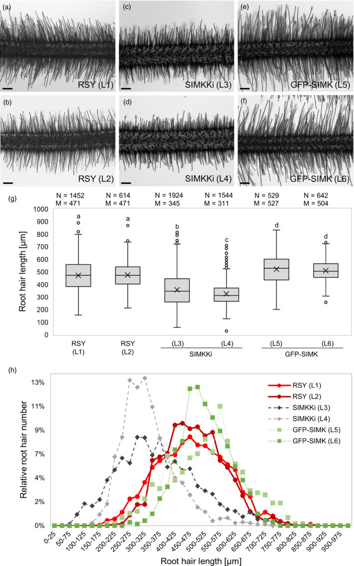
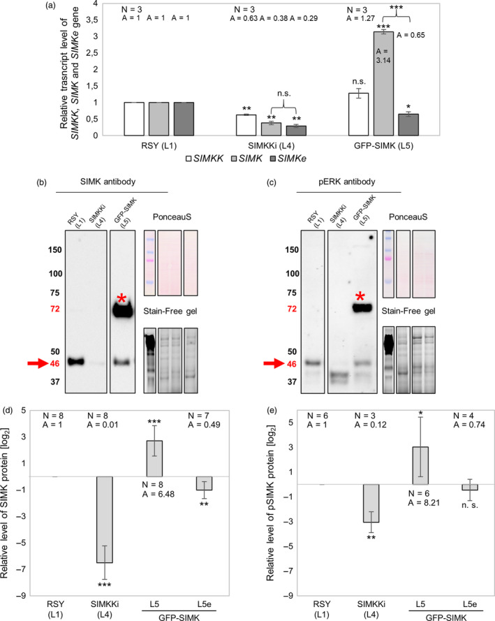
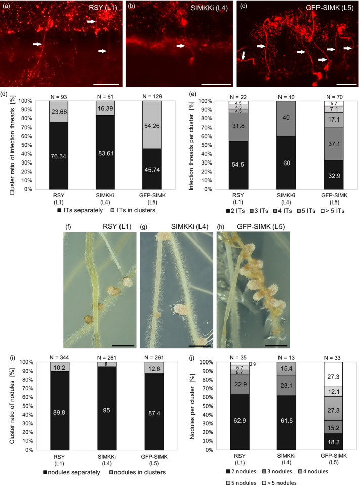
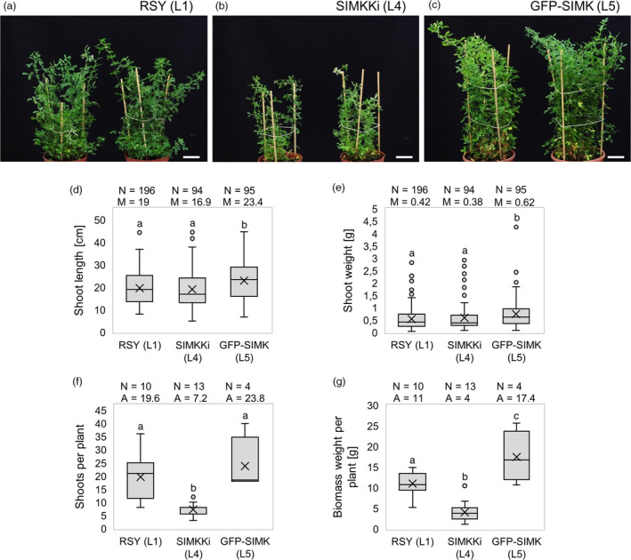
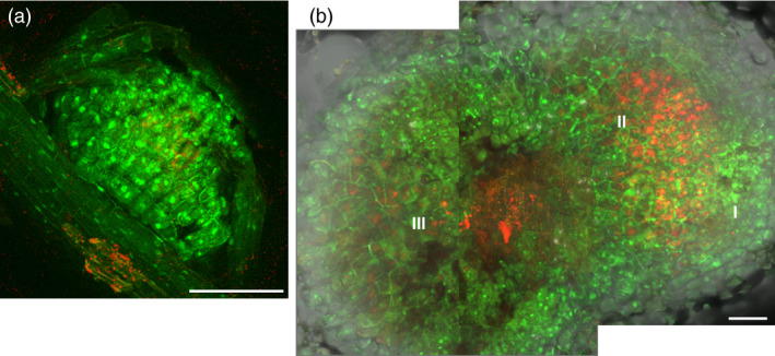
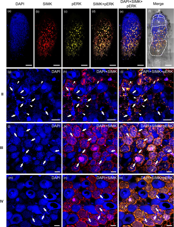
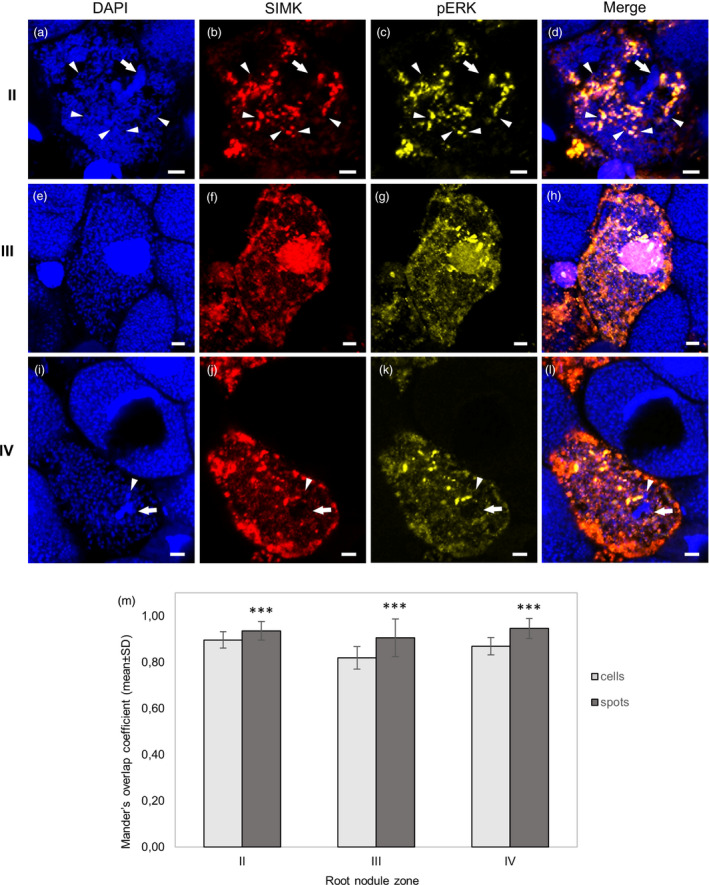
Nitrogen-fixing rhizobia and legumes have developed complex mutualistic mechanism that allows to convert atmospheric nitrogen into ammonia. Signalling by mitogen-activated protein kinases (MAPKs) seems to be involved in this symbiotic interaction. Previously, we reported that stress-induced MAPK (SIMK) shows predominantly nuclear localization in alfalfa root epidermal cells. Nevertheless, SIMK is activated and relocalized to the tips of growing root hairs during their development. SIMK kinase (SIMKK) is a well-known upstream activator of SIMK. Here, we characterized production parameters of transgenic alfalfa plants with genetically manipulated SIMK after infection with Sinorhizobium meliloti. SIMKK RNAi lines, causing strong downregulation of both SIMKK and SIMK, showed reduced root hair growth and lower capacity to form infection threads and nodules. In contrast, constitutive overexpression of GFP-tagged SIMK promoted root hair growth as well as infection thread and nodule clustering. Moreover, SIMKK and SIMK downregulation led to decrease, while overexpression of GFP-tagged SIMK led to increase of biomass in above-ground part of plants. These data suggest that genetic manipulations causing downregulation or overexpression of SIMK affect root hair, nodule and shoot formation patterns in alfalfa, and point to the new biotechnological potential of this MAPK.

Keywords: Medicago sativa; SIMK; SIMKK; infection thread; nodule; root hair.


Conflict of interest statement

The authors declare no competing financial interests. Correspondence and requests for materials should be addressed to J.Š. (jozef.samaj@upol.cz).

Figures

Figure 1
Figure 1
Root hair phenotypes in alfalfa RSY, SIMKK RNAi (SIMKKi) lines and lines overexpressing GFP‐SIMK. (a,b) Representative images of root hair phenotypes of plants from two independent lines (L1, L2) of control wild‐type RSY, (c,d) two independent transgenic lines with SIMKK RNAi construct (SIMKKi L3, L4) and (e,f) two independent transgenic lines expressing 35S::GFP:SIMK in wild‐type RSY background (GFP‐SIMK L5, L6). (g) Box plot graph depicting comparison in root hair length in indicated lines, number of observations N and median value M. Statistics was calculated in SigmaPlot11.0 using Kruskal–Wallis one‐way analysis of variance on ranks (Dunn's method) and is based on N = 529–1924. The numbers of root hairs observed were 1452 (RSY L1), 614 (RSY L2), 1924 (SIMKKi L3), 1544 (SIMKKi L4), 529 (GFP‐SIMK L5) and 642 (GFP‐SIMK L6). Different lower case letters indicate statistical significance between treatments (P < 0.05). (h) Relative distribution of root hair lengths in indicated alfalfa lines. Normalized root hair number was evaluated using 25 µm intervals distribution. Transgenic lines show different distribution pattern of root hair lengths as compared to RSY wild‐type lines. Scale bar: (a–f) 200 µm.
Figure 2
Figure 2
Expression analysis of SIMKK and SIMK genes by quantitative real‐time (qRT‐PCR) and immunoblotting analysis of total endogenous SIMK, active endogenous SIMK and both total and active GFP‐SIMK. (a) Deregulated transcript levels of SIMKK, total (endogenous native SIMKe + GFP‐tagged) SIMK and endogenous native SIMKe gene in SIMKKi L4 and GFP‐SIMK L5 transgenic lines of alfalfa. (b) Western blot detection of SIMK and GFP‐SIMK bands using SIMK antibody and (c) detection of active amount of respective proteins pSIMK and GFP‐pSIMK bands using pERK antibody in root tissue of control and transgenic alfalfa plants of SIMKKi (L4) and expressing 35S::GFP:SIMK (L5). Arrows point to the 46 kDa which corresponds to (b) endogenous SIMK and (c) endogenous pSIMK, while asterisks show bands around 72 kDa which corresponds to (b) GFP‐SIMK and (c) GFP‐pSIMK. (d,e) Log2 graphs depicting comparison of protein levels in respective lines (SIMKKi L4, GFP‐SIMK L5) relative to RSY L1, number of observations N and average value A (presented as inversed log2 values). GFP‐SIMK L5e refer to endogenous level of protein, while GFP‐SIMK L5 refers to GFP‐SIMK level. (d) Relative SIMK protein level in roots of control and transgenic plants (RSY L1, SIMKKi L4, GFP‐SIMK L5). (e) Relative pSIMK protein level in roots of control and transgenic plants (RSY L1, SIMKKi L4, GFP‐SIMK L5). (a,d,e) Statistics was calculated in Microsoft Excel using t‐test and is based on N = 3–8. Error bars show ± SD. Asterisks indicate statistical significance between treatments, *P < 0.05, **P < 0.01, ***P < 0.001, n. s. indicates no statistical significance.
Figure 3
Figure 3
Infection thread and nodule formation in alfalfa roots inoculated with Sinorhizobium meliloti‐mRFP. (a–c) Overview of the infection threads containing S. meliloti with mRFP (white arrows) in roots of (a) wild‐type RSY line L1, (b) in transgenic SIMKKi line L4 and (c) in transgenic GFP‐SIMK line L5 at 10 dpi. (d) Ratio of individual/clustered infection threads (in %) at 10 dpi. (e) Number of infection threads per cluster (in %) at 10 dpi. (f–h) Representative images of root nodules formed in respective alfalfa lines, (f) control RSY line L1, (g) SIMKKi line L4 and (h) GFP‐SIMK line L5 inoculated with S. meliloti‐mRFP 15 dpi on Fåhreus medium. (i) Ratio of individual/clustered nodules (in %) at 15 dpi. (j) Number of nodules per cluster (in %) at 15 dpi. N = number of observations. Scale bar: (a–c) 100 µm, (f–h) 1 cm. Dpi = day post‐inoculation.
Figure 4
Figure 4
Shoot biomass production in transgenic alfalfa plants grown in vivo. (a–c) Representative images of above‐ground parts of mature plants grown in pots in control RSY L1 (a), SIMKKi L4 (b) and GFP‐SIMK L5 (c). Regrown plants were documented 60 days after cutting the shoots. (d) Box plot graph depicting comparison in shoot length of indicated lines, number of observations N and median value M. (e) Box plot graph depicting comparison in shoot weight of indicated lines, number of observations N and median value M. (f) Box blot graph depicting comparison in number of shoots per plant of indicated lines, number of observations N and average value A. (g) Box plot graph depicting comparison in biomass weight per plant of indicated lines, number of observations N and average value A. Statistics was calculated in SigmaPlot11.0 using Kruskal–Wallis one‐way analysis of variance on ranks (Dunn's method) (d, e), or using one‐way analysis of variance (Holm–Sidak method) (f, g) and is based on (d, e) N = 94–196 and (f, g) N = 4–13. Different lower case letters indicate statistical significance between treatments (P < 0.05). Scale bar: (a–c) 4 cm.
Figure 5
Figure 5
Localization of GFP‐SIMK in alfalfa root nodules. Examples of nodule (a) at the early stage of development and (b) at the late stage of development observed by CLSM. Localization of fused GFP‐SIMK protein (green) in root nodules induced after inoculation with S. meliloti‐mRFP (red) on plants of GFP‐SIMK line L5 at 10 dpi (a) and 20 dpi (b). A composite image of two consequential frames is shown in (b). Tissue organization of the late nodule: I, meristematic zone; II, infection and differentiation zone; III, symbiotic zone. Scale bar: (a) 100 µm; (b) 200 µm.
Figure 6
Figure 6
Overview of SIMK localization and distribution in root nodules induced after Sinorhizobium meliloti inoculation of alfalfa control RSY L1 plants using immunofluorescence localization microscopy. (a–f) Overview of the representative root nodule. This overview was mounted as a composite image out of eight consequential frames. (a) Hand‐sectioned root nodules were stained for DNA using DAPI, and (b) immunostained for SIMK using anti‐AtMPK6 antibody and (c) for phosphorylated MAPKs using phospho‐specific pERK 44/42 antibody. (d) Overlay of SIMK and phosphorylated MAPKs, and (e) distribution of cell nuclei in SIMK‐phosphorylated MAPKs overlay. (f) Bright‐field image of the same nodule with overlaid fluorescence channels schematically depicts distribution of individual developmental zones, namely meristematic (I), infection (II), symbiotic (III) and senescent (IV) zones. (g‐o) Representative images of cells from different nodule developmental zones: (II, g–i) the infection zone, (III, j–l) the symbiotic zone and (IV, m–o) the senescent zone. (g,j,m) Blue channels represent DAPI staining, (h,k,n) red channels (overlaid with DAPI channel) represent SIMK immunolocalization, and (i,l,o) yellow channels (overlaid with DAPI channel) represent colocalization (in yellow colour) of SIMK with phosphorylated MAPKs. Note specific staining of bacteria with DAPI. Infection threads are pointed by arrows, and releases of bacteria from branched infection threads in the form of infection droplets are pointed by arrowheads. Scale bars: (a–f) 10 mm, (g–o) 20 µm.
Figure 7
Figure 7
Detailed SIMK localization in root nodule cells induced by Sinorhizobium meliloti on alfalfa control RSY L1 plants using immunofluorescence localization microscopy. (a–d) Cell of the infection zone (II) with branched infection thread (arrow) during the release of bacteria in the form of infection droplets (arrowheads). (e–h) Cells of the symbiotic zone (III) with developed bacteroids. (i–l) Cells of the senescent zone (IV) with developed bacteroids. (a,e,i) Nuclei and bacteria are stained with DAPI, (b,f,j) SIMK (in red) is immunostained with anti‐AtMPK6 antibody and (c,g,k) phosphorylated MAPKs (in yellow) are immunostained with phospho‐specific pERK 44/42 antibody. (d,h,l) Overlay of DAPI, SIMK and phosphorylated MAPKs. Note specific staining of bacteria with DAPI. Infection threads are pointed by arrows, and releases of bacteria from branched infection threads in the form of infection droplets are pointed by arrowheads. (m) Averaged Mander’s overlap coefficients from quantitative colocalization of SIMK with phosphorylated MAPKs evaluated in defined ROIs in each zone (cells), and in particular subcellular structures (spots) of the infection zone (II, N = 23 ROIs in cells and N = 84 in spots), the symbiotic zone (III, N = 23 ROIs in cells and N = 83 in spots) and the senescent zone (IV, N = 15 ROIs in cells and N = 32 in spots). Quantitative colocalization analysis is presented in Figures S8–S13. Statistics was calculated in Microsoft Excel using t‐test. Error bars show ± SD. Asterisks indicate statistical significance between treatments (***P < 0.001). Scale bar: (a–l) 5 µm.

Similar articles

Cited by

References

    1. Aung, B. , Gruber, M.Y. , Amyot, L. , Omari, K. , Bertrand, A. and Hannoufa, A. (2015) Micro RNA 156 as a promising tool for alfalfa improvement. Plant Biotechnol. J. 13, 779–790. - PubMed
    1. Baluška, F. , Ovečka, M. and Hirt, H. (2000a) Salt stress induces changes in amounts and localization of the mitogen‐activated protein kinase SIMK in alfalfa roots. Protoplasma, 212, 262–267.
    1. Baluška, F. , Salaj, J. , Mathur, J. , Braun, M. , Jasper, F. , Šamaj, J. , Chua, N. H. et al. (2000b) Root hair formation: F‐actin‐dependent tip growth is initiated by local assembly of profilin‐supported F‐actin meshworks accumulated within expansin‐enriched bulges. Dev. Biol. 227, 618–632. - PubMed
    1. Bekešová, S. , Komis, G. , Křenek, P. , Vyplelová, P. , Ovečka, M. , Luptovčiak, I. and Šamaj, J. (2015) Monitoring protein phosphorylation by acrylamide pendant Phos‐Tag™ in various plants. Front. Plant Sci. 6, 336. - PMC - PubMed
    1. Biazzi, E. , Nazzicari, N. , Pecetti, L. , Brummer, E.C. , Palmonari, A. , Tava, A. and Annicchiarico, P. (2017) Genome‐wide association mapping and genomic selection for alfalfa (Medicago sativa) forage quality traits. PLoS One, 12, e0169234. - PMC - PubMed

Publication types

Substances

LinkOut - more resources