. 2020 Dec;10(12):2510-2531.
doi: 10.1002/2211-5463.13016. Epub 2020 Nov 13.

The role of the cytoskeletal proteins MreB and FtsZ in multicellular cyanobacteria

Affiliations

The role of the cytoskeletal proteins MreB and FtsZ in multicellular cyanobacteria

Benjamin L Springstein et al. FEBS Open Bio. 2020 Dec.

Abstract

Multiseriate and true-branching cyanobacteria are at the peak of prokaryotic morphological complexity. However, little is known about the mechanisms governing multiplanar cell division and morphogenesis. Here, we study the function of the prokaryotic cytoskeletal proteins, MreB and FtsZ in Fischerella muscicola PCC 7414 and Chlorogloeopsis fritschii PCC 6912. Vancomycin and HADA labeling revealed a mixed apical, septal, and lateral trichome growth mode in F. muscicola, whereas C. fritschii exhibits septal growth. In all morphotypes from both species, MreB forms either linear filaments or filamentous strings and can interact with FtsZ. Furthermore, multiplanar cell division in F. muscicola likely depends on FtsZ dosage. Our results lay the groundwork for future studies on cytoskeletal proteins in morphologically complex cyanobacteria.

Keywords: Cyanobacteria; FtsZ; MreB; Stigonematales; cytoskeleton; morphogenesis.


Conflict of interest statement

The authors declare no conflict of interest.

Figures

Fig. 1
Fig. 1
Presence of ftsZ and mreB homologs in cyanobacteria. (A) Illustration depicting the cellular morphologies and life stages ofF. muscicola, C. fritschii, and Anabaena wild‐type trichomes. (B) Depiction of representative cyanobacterial species with different cell shapes and morphologies and from different habitats. The complete dataset is shown in File S1. Species, where homologous genes to ftsZ, ftsQ, mreB, mreC, and mreD were detected, are marked by a green square. Organisms, where a homolog was not found, are marked by a gray square.
Fig. 2
Fig. 2
Fluorescent vancomycin labeling reveals different growth modes in multicellular cyanobacteria. (A–G) Bright‐field and merged chlorophyll autofluorescence (red) and BODIPY ®FL Vancomycin (Van‐FL) fluorescence micrographs of (A–C) F. muscicola, (D–F)C. fritschii, and (G) Anabaena cells stained with 5 µg·mL−1Van‐FL. Micrographs indicate different growth stages of the respective cyanobacterium: (A upper image) hormogonia, (A lower image) hormogonia in the transition phase to young linear trichome. Apical staining was observed in 123 apical cells (39%) while 191 showed septal staining only (61%),n = 314. (B) young branches, (C, F) heterocysts, (D) hormogonium (as indicated by the linear growth mode, which is restricted to the early growth stages (i.e. hormogonia) ofC. fritschii), (E) multiseriate trichome, and (G) mature trichome. White arrows indicate heterocysts. (B) and (B inlay) additionally show Van‐FL staining pattern at the branching points of newly formed lateral branches. Van‐FL staining patterns were consistent among three independent experiments. Scale bars: 10 µm.
Fig. 3
Fig. 3
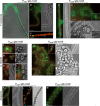
Localization of GFP‐MreB in F. muscicola, C. fritschii, and Anabaena. Bright‐field and merged chlorophyll autofluorescence (red) and GFP fluorescence micrographs of (A–E) F. muscicola, (F–J) C. fritschii, or (K–M) Anabaena expressing gfp‐mreB from (A–C, F–I, K–L) PpetE or from (D, E, J, M) PmreB. Figures show (A) mature trichome with nascent hormogonium, (B, E–G, J, K–M) mature trichomes and (D, H, I, J inlay) hormogonia. A heterocyst is marked with an orange triangle. Blue triangles indicate longitudinal GFP‐MreB filaments that appear to traverse the cells along the growth axis. Note: Anabaenaexpressing gfp‐mreBfrom PpetEshows several small filaments throughout the cells when grown on BG11 medium, while polar GFP‐MreB plugs are only observed upon transfer to BG110(BG11 without combined nitrogen), which we found to seemingly increase PpetE‐driven expression, thus indicating that the GFP spots seen in (L) could be inclusion body‐like aggregates. Scale bars: (A–G, J–M) 10 µm or (H, I) 5 µm.
Fig. 4
Fig. 4
Interaction of FtsZ and MreB in multicellular cyanobacteria. (A) Beta‐galactosidase assays ofEscherichia colicells co‐expressing indicated translational fusion constructs of all possible pairwise combinations offtsZwithmreB. Quantity values are given in Miller units per milligram LacZ of the mean results from three independent colonies. Error bars indicate standard deviations (n = 3). Neg: pKNT25 plasmid carryingftsZco‐transformed with empty pUT18C. Pos: Zip/Zip control. The value indicated with **** is significantly different from the negative control (****P < 0.0001; Dunnett's multiple comparison test and one‐way ANOVA). (B) Anti‐FtsZSubsVand anti‐GFP western blots from anti‐GFP co‐IP's from two individual biological samples ofF. muscicola,C. fritschii, andAnabaenacell‐free extracts expressinggfp‐mreBfrom PmreBor WT control cells. Experiments were performed twice, each time using two independent biological replicates for each sample type (i.e. WT and GFP‐tagged MreB expressing strain). Input samples were taken before addition of anti‐GFP antibodies.
Fig. 5
Fig. 5
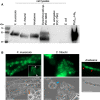
Assessment of new Anti‐FtsZSubsVantibody. (A) Western blot analysis of cell‐free lysates fromF. muscicola,C. fritschii,Anabaena,Synechocystis,Synechococcus, andEscherichia coliusing anti‐FtsZSubsVantibody (raised againstF. muscicolaandC. fritschiiFtsZ). As a control, 1 µg purified FtsZFm‐His was included. (B) Immunolocalization of FtsZ inF. muscicola,C. fritschii, andAnabaenausing an anti‐FtsZSubsVantibody. White triangles show Z‐rings in branching cells. The orange arrow indicates a heterocyst. The blue triangle points to a matureC. fritschiicell which was not permeabilized. Red triangles indicate septal localization of FtsZ inAnabaena. Results shown in here are representative figures from three independent experiments. Scale bars: 10 µm.
Fig. 6
Fig. 6
Fischerella muscicolaandAnabaenaare sensitive to excess FtsZ levels. (A) Micrographs of mating filters showing growth ofF. muscicolaWT colonies andF. muscicolacolonies after transformation with pRL25C plasmids carrying PpetE::ftsZ‐gfp, PglnA::gfp‐ftsZor PftsZ::ftsZ‐gfp. (B) Bright‐field micrograph showing morphological alterations (i.e. swelling) ofF. muscicolacells expressingftsZ‐gfpfrom PftsZ. (C–E) Merged chlorophyll autofluorescence (red) and GFP fluorescence and bright‐field micrographs of (C)F. muscicola, (D)Anabaenaor (E)C. fritschiiexpressingftsZ‐gfporgfp‐ftsZfrom PftsZ, PglnAor PpetE. PpetE‐driven expression offtsZ‐gfpinF. muscicolaandAnabaenawas additionally increased using 0.2 µmCuSO4. White arrows indicate trichomes with multiseriate trichome growth. Blue arrows mark dim Z‐ring formations. Scale bars: (C–E) 10 µm.
Fig. 7
Fig. 7
High rate of proteolytic degradation of FtsZ in subsection V cyanobacteria. (A) Anti‐FtsZSubsVwestern blots of cell‐free extracts ofF. muscicola,C. fritschii, andAnabaenaincubated at 37 °C (F. muscicolaandC. fritschii) or 30 °C (Anabaena) for 0, 60, 120, or 180 min supplemented or not (w/o) with protease inhibitor cocktails (PIC) and EDTA/EGTA. Cells were either lysed in cyanobacterial lysis buffer (CLB) or in CLB supplemented with 10 mmMgCl2. EDTA, and EGTA were supplemented as 5 mmeach, except in cases of presence of 10 mmMgCl2, where 7.5 mmEDTA and EGTA were included. (B) Purified FtsZFm‐His (1 µg per sampling time point) was incubated for the indicated time points in cell‐free extracts ofF. muscicola,C. fritschii, orAnabaenasupplemented or not (w/o) with PIC and EDTA/EGTA. Degradation of FtsZFm‐His was detected using an anti‐His antibody. (A, B) As a loading and specific‐degradation control, levels of RbcL were detected using an anti‐RbcL antibody. Images are representative western blots from duplicate experiments.

References

    1. Typas A, Banzhaf M, Gross CA and Vollmer W (2012) From the regulation of peptidoglycan synthesis to bacterial growth and morphology. Nat Rev Microbiol 10, 123. - PMC - PubMed
    1. Kysela DT, Randich AM, Caccamo PD and Brun YV (2016) Diversity takes shape: understanding the mechanistic and adaptive basis of bacterial morphology. PLoS Biol 14, e1002565. - PMC - PubMed
    1. Kruse T, Bork‐Jensen J and Gerdes K (2005) The morphogenetic MreBCD proteins of Escherichia coli form an essential membrane‐bound complex. Mol Microbiol 55, 78–89. - PubMed
    1. Domínguez‐Escobar J, Chastanet A, Crevenna AH, Fromion V, Wedlich‐Söldner R and Carballido‐López R (2011) Processive movement of MreB‐associated cell wall biosynthetic complexes in bacteria. Science 333, 225–228. - PubMed
    1. Garner EC, Bernard R, Wang W, Zhuang X, Rudner DZ and Mitchison T (2011) Circumferential motions of the cell wall synthesis machinery drive cytoskeletal dynamics in B. subtilis . Science 333, 222–225. - PMC - PubMed

Publication types

MeSH terms

Supplementary concepts