

Integration of Simultaneous Resting-State Electroencephalography, Functional Magnetic Resonance Imaging, and Eye-Tracker Methods to Determine and Verify Electroencephalography Vigilance Measure
- PMID: 33112650
- PMCID: PMC7757617
- DOI: 10.1089/brain.2019.0731
Integration of Simultaneous Resting-State Electroencephalography, Functional Magnetic Resonance Imaging, and Eye-Tracker Methods to Determine and Verify Electroencephalography Vigilance Measure
Abstract
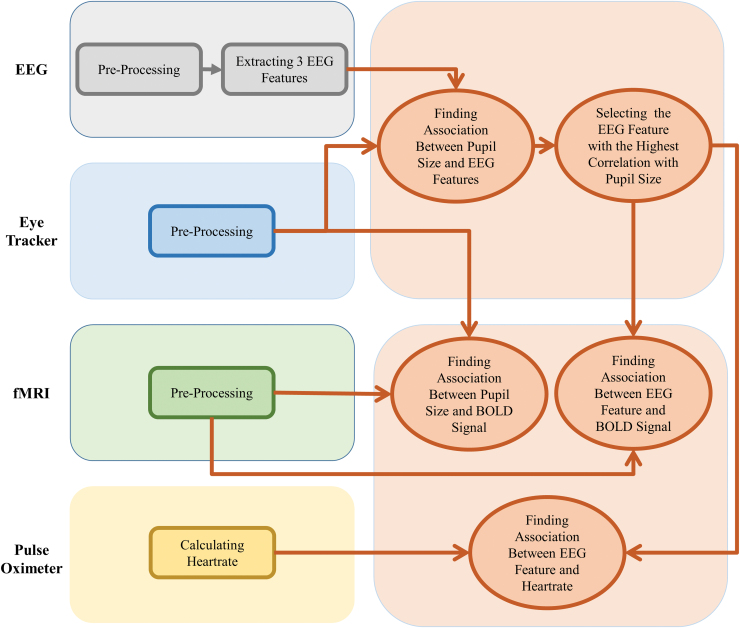
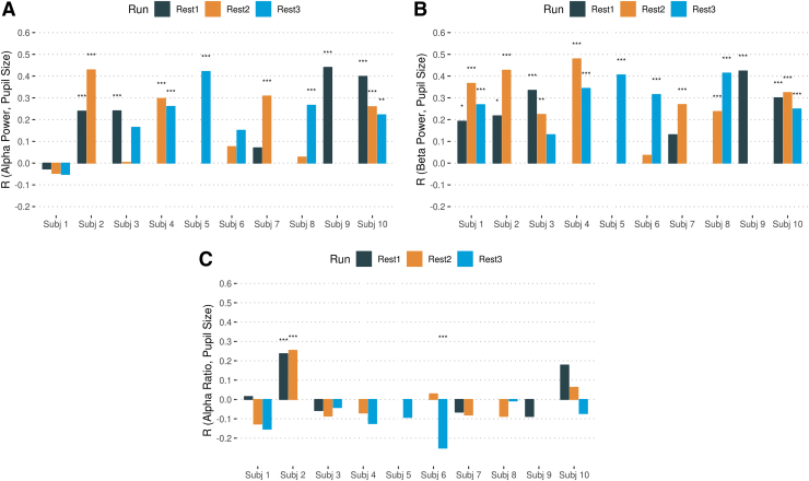
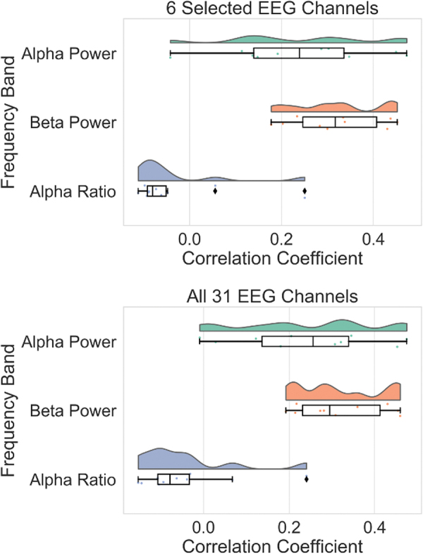
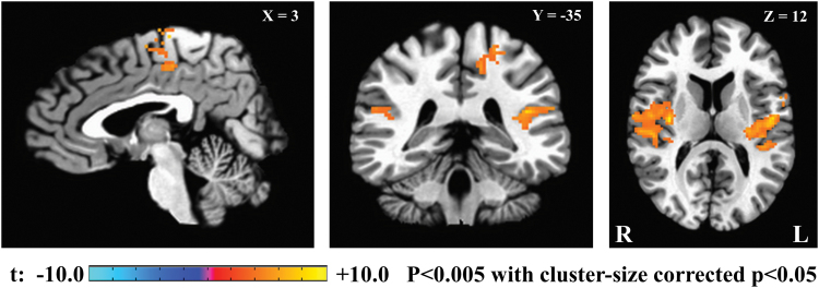
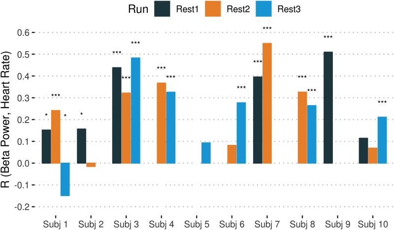
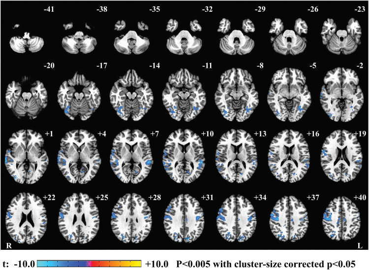
Background/Introduction: Concurrent electroencephalography and resting-state functional magnetic resonance imaging (rsfMRI) have been widely used for studying the (presumably) awake and alert human brain with high temporal/spatial resolution. Although rsfMRI scans are typically collected while individuals are instructed to focus their eyes on a fixated cross, objective and verified experimental measures to quantify degree of vigilance are not readily available. Electroencephalography (EEG) is the modality extensively used for estimating vigilance, especially during eyes-closed resting state. However, pupil size measured using an eye-tracker device could provide an indirect index of vigilance. Methods: Three 12-min resting scans (eyes open, fixating on the cross) were collected from 10 healthy control participants. We simultaneously collected EEG, fMRI, physiological, and eye-tracker data and investigated the correlation between EEG features, pupil size, and heart rate. Furthermore, we used pupil size and EEG features as regressors to find their correlations with blood-oxygen-level-dependent fMRI measures. Results: EEG frontal and occipital beta power (FOBP) correlates with pupil size changes, an indirect index for locus coeruleus activity implicated in vigilance regulation (r = 0.306, p < 0.001). Moreover, FOBP also correlated with heart rate (r = 0.255, p < 0.001), as well as several brain regions in the anticorrelated network, including the bilateral insula and inferior parietal lobule. Discussion: In this study, we investigated whether simultaneous EEG-fMRI combined with eye-tracker measurements can be used to determine EEG signal feature associated with vigilance measures during eyes-open rsfMRI. Our results support the conclusion that FOBP is an objective measure of vigilance in healthy human subjects. Impact statement We revealed an association between electroencephalography frontal and occipital beta power (FOBP) and pupil size changes during an eyes-open resting state, which supports the conclusion that FOBP could serve as an objective measure of vigilance in healthy human subjects. The results were validated by using simultaneously recorded heart rate and functional magnetic resonance imaging (fMRI). Interestingly, independently verified heart rate changes can also provide an easy-to-determine measure of vigilance during resting-state fMRI. These findings have important implications for an analysis and interpretation of dynamic resting-state fMRI connectivity studies in health and disease.
Keywords: EEG; eye tracker; heart rate; pupillometry; resting-state fMRI; vigilance.
Conflict of interest statement
The authors declare no competing financial interests.
Figures
References
-
- Allen PJ, Josephs O, Turner R. 2000. A method for removing imaging artifact from continuous EEG recorded during functional MRI. Neuroimage 12:230–239 - PubMed
-
- Allen PJ, Polizzi G, Krakow K, Fish DR, Lemieux L. 1998. Identification of EEG events in the MR scanner: the problem of pulse artifact and a method for its subtraction. Neuroimage 8:229–239 - PubMed
-
- Barry RJ, Clarke AR, Johnstone SJ, Magee CA, Rushby JA. 2007. EEG differences between eyes-closed and eyes-open resting conditions. Clin Neurophysiol 118:2765–2773 - PubMed
-
- Bell AJ, Sejnowski TJ. 1995. An information-maximization approach to blind separation and blind deconvolution. Neural Comput 7:1129–1159 - PubMed
-
- Belyavin A, Wright NA. 1987. Changes in electrical activity of the brain with vigilance. Electroencephalogr Clin Neurophysiol 66:137–144 - PubMed
Publication types
MeSH terms
LinkOut - more resources
Full Text Sources
Medical
