Cdc1p is a Golgi-localized glycosylphosphatidylinositol-anchored protein remodelase
- PMID: 33112703
- PMCID: PMC7927193
- DOI: 10.1091/mbc.E20-08-0539
Cdc1p is a Golgi-localized glycosylphosphatidylinositol-anchored protein remodelase
Abstract
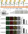
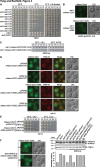
Glycosylphosphatidylinositol-anchored proteins (GPI-APs) undergo extensive posttranslational modifications and remodeling, including the addition and subsequent removal of phosphoethanolamine (EtNP) from mannose 1 (Man1) and mannose 2 (Man2) of the glycan moiety. Removal of EtNP from Man1 is catalyzed by Cdc1p, an event that has previously been considered to occur in the endoplasmic reticulum (ER). We establish that Cdc1p is in fact a cis/medial Golgi membrane protein that relies on the COPI coatomer for its retention in this organelle. We also determine that Cdc1p does not cycle between the Golgi and the ER, and consistent with this finding, when expressed at endogenous levels ER-localized Cdc1p-HDEL is unable to support the growth of cdc1Δ cells. Our cdc1 temperature-sensitive alleles are defective in the transport of a prototypical GPI-AP-Gas1p to the cell surface, a finding we posit reveals a novel Golgi-localized quality control warrant. Thus, yeast cells scrutinize GPI-APs in the ER and also in the Golgi, where removal of EtNP from Man2 (via Ted1p in the ER) and from Man1 (by Cdc1p in the Golgi) functions as a quality assurance signal.
Figures


Similar articles
-
Remodeling-defective GPI-anchored proteins on the plasma membrane activate the spindle assembly checkpoint.Cell Rep. 2021 Dec 28;37(13):110120. doi: 10.1016/j.celrep.2021.110120. Cell Rep. 2021. PMID: 34965437
-
Two endoplasmic reticulum (ER) membrane proteins that facilitate ER-to-Golgi transport of glycosylphosphatidylinositol-anchored proteins.Mol Biol Cell. 1999 Apr;10(4):1043-59. doi: 10.1091/mbc.10.4.1043. Mol Biol Cell. 1999. PMID: 10198056 Free PMC article.
-
Cdc1 removes the ethanolamine phosphate of the first mannose of GPI anchors and thereby facilitates the integration of GPI proteins into the yeast cell wall.Mol Biol Cell. 2014 Nov 1;25(21):3375-88. doi: 10.1091/mbc.E14-06-1033. Epub 2014 Aug 27. Mol Biol Cell. 2014. PMID: 25165136 Free PMC article.
-
Transport of glycosylphosphatidylinositol-anchored proteins from the endoplasmic reticulum.Biochim Biophys Acta. 2013 Nov;1833(11):2473-8. doi: 10.1016/j.bbamcr.2013.01.027. Epub 2013 Feb 1. Biochim Biophys Acta. 2013. PMID: 23380706 Review.
-
Trafficking of glycosylphosphatidylinositol anchored proteins from the endoplasmic reticulum to the cell surface.J Lipid Res. 2016 Mar;57(3):352-60. doi: 10.1194/jlr.R062760. Epub 2015 Oct 8. J Lipid Res. 2016. PMID: 26450970 Free PMC article. Review.
Cited by
-
Potential Physiological Relevance of ERAD to the Biosynthesis of GPI-Anchored Proteins in Yeast.Int J Mol Sci. 2021 Jan 21;22(3):1061. doi: 10.3390/ijms22031061. Int J Mol Sci. 2021. PMID: 33494405 Free PMC article. Review.
-
Unremodeled GPI-anchored proteins at the plasma membrane trigger aberrant endocytosis.Life Sci Alliance. 2024 Nov 22;8(2):e202402941. doi: 10.26508/lsa.202402941. Print 2025 Feb. Life Sci Alliance. 2024. PMID: 39578075 Free PMC article.
-
Structure and Function of the Glycosylphosphatidylinositol Transamidase, a Transmembrane Complex Catalyzing GPI Anchoring of Proteins.Subcell Biochem. 2024;104:425-458. doi: 10.1007/978-3-031-58843-3_16. Subcell Biochem. 2024. PMID: 38963495 Review.
-
Recent research progress in glycosylphosphatidylinositol-anchored protein biosynthesis, chemical/chemoenzymatic synthesis, and interaction with the cell membrane.Curr Opin Chem Biol. 2024 Feb;78:102421. doi: 10.1016/j.cbpa.2023.102421. Epub 2024 Jan 4. Curr Opin Chem Biol. 2024. PMID: 38181647 Free PMC article. Review.
-
To each its own: Mechanisms of cross-talk between GPI biosynthesis and cAMP-PKA signaling in Candida albicans versus Saccharomyces cerevisiae.J Biol Chem. 2024 Jul;300(7):107444. doi: 10.1016/j.jbc.2024.107444. Epub 2024 Jun 4. J Biol Chem. 2024. PMID: 38838772 Free PMC article. Review.
References
-
- Benachour A, Sipos G, Flury I, Reggiori F, Canivenc-Gansel E, Vionnet C, Conzelmann A, Benghezal M (1999). Deletion of GPI7, a yeast gene required for addition of a side chain to the glycosylphosphatidylinositol (GPI) core structure, affects GPI protein transport, remodeling, and cell wall integrity. J Biol Chem 274, 15251–15261. - PubMed
-
- Caro LH, Tettelin H, Vossen JH, Ram AF, van den Ende H, Klis FM (1997). In silicio identification of glycosyl-phosphatidylinositol-anchored plasma-membrane and cell wall proteins of Saccharomyces cerevisiae. Yeast 13, 1477–1489. - PubMed
-
- Castillon GA, Watanabe R, Taylor M, Schwabe TME, Riezman H (2009). Concentration of GPI-anchored proteins upon ER exit in yeast. Traffic 10, 186–200. - PubMed
-
- Fujita M, Kinoshita T (2012). GPI-anchor remodeling: potential functions of GPI-anchors in intracellular trafficking and membrane dynamics. Biochim Biophys Acta 1821, 1050–1058. - PubMed
Publication types
MeSH terms
Substances
LinkOut - more resources
Full Text Sources
Molecular Biology Databases
Miscellaneous

