. 2020 Dec;27(12):671-683.
doi: 10.1530/ERC-20-0258.

Amphiregulin retains ERα expression in acquired aromatase inhibitor resistant breast cancer cells

Affiliations

Amphiregulin retains ERα expression in acquired aromatase inhibitor resistant breast cancer cells

Yuanzhong Wang et al. Endocr Relat Cancer. 2020 Dec.

Abstract

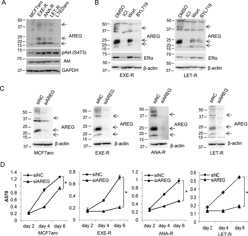
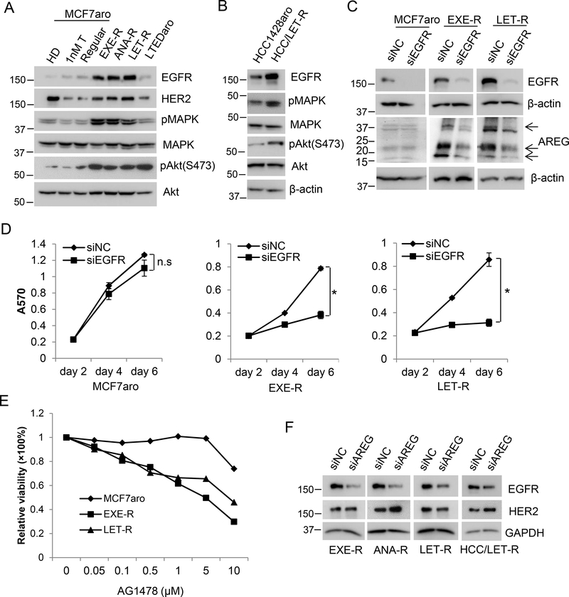
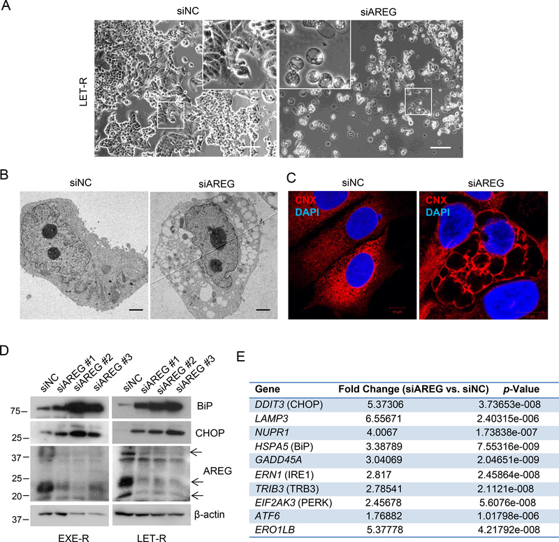
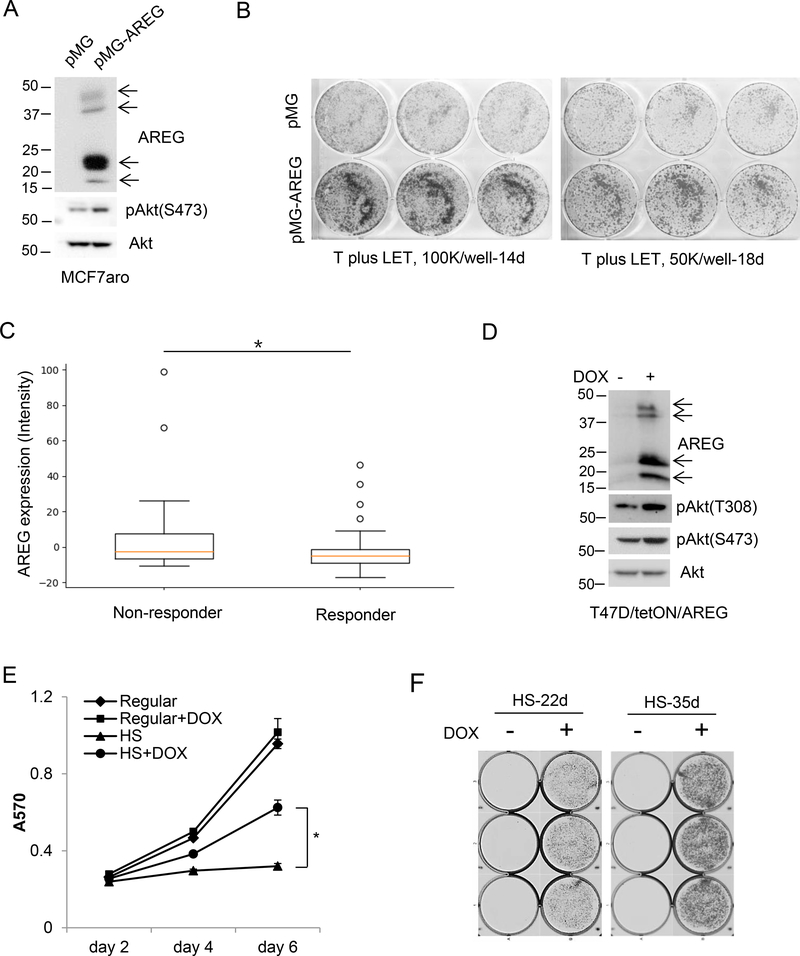
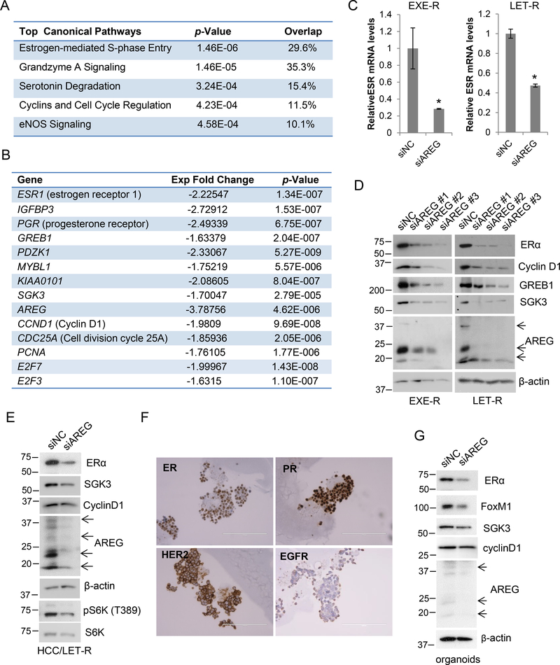
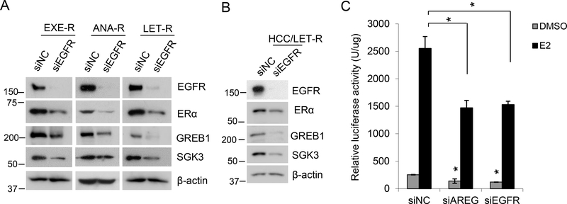
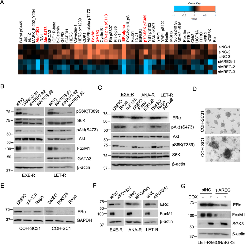
Acquired resistance to aromatase inhibitors (AIs) is a significant clinical issue in endocrine therapy for estrogen receptor (ER) positive breast cancer which accounts for the majority of breast cancer. Despite estrogen production being suppressed, ERα signaling remains active and plays a key role in most AI-resistant breast tumors. Here, we found that amphiregulin (AREG), an ERα transcriptional target and EGF receptor (EGFR) ligand, is crucial for maintaining ERα expression and signaling in acquired AI-resistant breast cancer cells. AREG was deregulated and critical for cell viability in ER+ AI-resistant breast cancer cells, and ectopic expression of AREG in hormone responsive breast cancer cells promoted endocrine resistance. RNA-sequencing and reverse phase protein array analyses revealed that AREG maintains ERα expression and signaling by activation of PI3K/Akt/mTOR signaling and upregulation of forkhead box M1 (FOXM1) and serum- and glucocorticoid-inducible kinase 3 (SGK3) expression. Our study uncovers a previously unappreciated role of AREG in maintaining ERα expression and signaling, and establishes the AREG-ERα crosstalk as a driver of acquired AI resistance in breast cancer.

Keywords: FOXM1; SGK3; endocrine resistance; estrogen receptor; mTOR.


Conflict of interest statement

Conflict of interest: The authors declare that they have no conflicts of interest with the contents of this article.

Figures

Figure 1.
Figure 1.. AREG is deregulated and critical for cell viability in acquired AI-resistant cells.
(A) Western blotting analysis of AREG expression. All the cell lines were cultured in their normal growth media. (B) Western blotting analysis of EXE-R and LET-R cells after treatment with DMSO, 100nM ICI, 0.2μM Wortmannin, or 5μM BYL719 for 72h. (C, D) MCF7aro cells and AI-resistant cells were transfected with siRNA negative control or AREG siRNA. Cells were harvested for Western blotting analysis (C) at 72 h post-transfection or measured for cell viability by MTT assay (D) at the time indicated. *, p<0.05.
Figure 2.
Figure 2.. EGFR signaling is deregulated in acquired AI-resistant cells.
(A) Western blotting analysis of EGFR and the related proteins. MCF7aro cells were cultured in the normal growth medium (regular), or hormone deprived for 2 days (HD) and treated with 1nM testerosterone (T) for two days. All the other cell lines were cultured in their normal growth media. (B) Western blotting analysis of HCC1428aro cells and HCC1428aro/LET-R cells. Both cell lines were cultured in their normal growth medium. (C, D) MCF7aro, EXE-R, and LET-R cells were transfected with siRNA negative control or EGFR siRNA, respectively. Cells were harvested for Western blotting analysis (C) at 72 h post-transfection or measured for cell viability by MTT assay (D) at the time indicated. n.s: not significant; *, p<0.05. (E) The inhibition of AG1478 on MCF7aro cells and AI-resistant cells. MCF7aro, EXE-R and LET-R cells were treated with the indicated concentrations of AG1478 for 96h, and cell viability was measured by MTT. The inhibition was calculated compared to solvent treatment. (F) Western blotting analysis of four AI-resistant cell lines after transfection with siRNA negative control or AREG siRNA.
Figure 3.
Figure 3.. AREG is required for EnR homoestasis of acquired AI-resistant cells.
(A) Phase contrast microscopy of LET-R cells transfected with siRNA negative control or AREG siRNA for 72h. Scale bar: 50μm. (B) Electron microscopy of LET-R cells transfected with siRNA negative control or AREG siRNA for 72h. Scale bar: 2μm. (C) Immunofluorescence microscopy of LET-R cells transfected with siRNA negative control or AREG siRNA. After transfection for 72 h, cells were fixed and immunostained with anti-calnexin (CNX). After immunostaining, the cells were mounted in DAPI solution and imaged under a confocal microscope. DAPI stained nuclei blue. CNX was shown in red. Scale bar: 10μm. (D) Western blotting analysis of EXE-R and LET-R cells after transfection with siRNA negative control or three individual AREG siRNAs, respectively. (E) The list of EnR stress-related genes upregulated in AREG knockdown LET-R cells in RNA-Sequencing analysis.
Figure 4.
Figure 4.. Overexpression of AREG promotes endocrine therapy resistance.
(A) Western blotting analysis of MCF7aro cells stably transfected with empty vector pMG or AREG expressing vector. (B) MCF7aro/pMG and MCF7aro/pMG-AREG cells were seeded into 6-well plates at 100,000 cells/well or 50,000 cells/well, and cultured in hormone-stripped medium and treated with 1nM testosterone plus 200nM LET for 14 days and 18 days, respectively. Cells were fixed and stained with crystal violet. Images were taken using a scanner. (C) Plot of AREG expression vs. clinical response to LET in 75 breast tumors (23 nonresponders and 52 responders) from breast cancer dataset (GSE59515). The mean of AREG expression intensity was 7.413 in nonresponderand and −2.816 in responders. *, p=0.0222 by Student’s t test. (D) Western blotting analysis of T47D/tetON/AREG cells cultured in regular medium in the presence or absence of 100ng/ml DOX for 48h. (E) T47D/tetON/AREG cells were cultured in normal growth medium (regular) or hormone-stripped medium (HS) in the presence or absence of 100ng/ml DOX. Cell viability was measured by MTT assay. *, p<0.05. (F) T47D/tetON/AREG cells were seeded into 6-well plates at 50,000 cells/well, and cultured in hormone-stripped medium in the presence or absence of 100ng/ml DOX. After 22 days and 35 days, cells were fixed and stained with crystal violet. Image was captured by Cell3 iMager duos system.
Figure 5.
Figure 5.. AREG maintains ERα expression and signaling.
(A, B) RNA-Sequencing analysis of LET-R cells transfected with siRNA negative control or AREG siRNA. The top canonical pathways affected by knockdown of AREG analyzed by Ingenuity pathway analysis (IPA) were shown in (A). The expression changes of ESR1 (the gene coding ERα) and ERα-target genes as well as genes involving cell proliferation were shown in (B). (C) RT-qPCR analysis of ESR1 mRNA levels after transfection with siRNA negative control or AREG siRNA in EXE-R and LET-R cells. (D) Western blotting analysis of EXE-R and LET-R cells after transfected with siRNA negative control or three individual AREG siRNAs, respectively. (E) Western blotting analysis of HCC1428aro/LET-R cells transfected with siRNA negative control or the pooled AREG siRNA. (F) Immunohistochemistry staining of COH-SC31 organoids for detection of ERα, PR, HER2 and EGFR. Organoids were similar to the original tumor in expression of ERα, PR, HER2. (G) Western blotting analysis of COH-SC31 organoids transfected with siRNA negative control or AREG siRNA for 90h.
Figure 6.
Figure 6.. EGFR regulates ERα expression and signaling.
(A) Western blotting analysis of AI-resistant cells transfected with siRNA negative control or EGFR siRNA for 72h. (B) Western blotting analysis of HCC1428aro/LET-R cells transfected with siRNA negative control or EGFR siRNA for 72h. (C) Luciferase reporter assay for ERα transactivity. MCF7aro/ERE cells were cultured in hormone-deprived medium and transfected with siRNA negative control, AREG siRNA or EGFR siRNA. Meanwhile, cells were treated with or without 10nM E2. At 48h posttreatment, cells were harvested for luciferase activity assay. Luciferase activity was normalized to protein concentration. *, p<0.05 vs. siRNA NC, n=3.
Figure 7.
Figure 7.. AREG sustains ERα signaling by activation of PI3K/Akt/mTOR signaling and upregulation of FoxM1 and SGK3 expression.
(A) The partial heatmap of RPPA of LET-R cells transfected with siRNA negative control or AREG siRNA, which highlights most of downregulated proteins in AREG knockdown cells. The full heatmap was shown in supplementary Fig.7A. (B) Western blotting analysis of AI-resistant cells transfected with siRNA negative control or 3 individual AREG siRNAs, respectively. (C) Western blotting analysis of AI-resistant cells treated with 0.5μM rapamycin or 0.5μM INK128. (D) Light microscopy of the organoids generated from COH-SC31 and COH-SC1 PDXs. (E) Western blotting analysis of COH-SC31 and COH-SC1 organoids treated with 0.5μM rapamycin or 0.5μM INK128. (F) Western blotting analysis of AI-resistant cells transfected with siRNA negative control or FoxM1 siRNA. (G) Western blotting analysis of LET-R/TetON/SGK3 cells transfected with siRNA negative control or AREG siRNA in the presence or absence of doxycycline.

Similar articles

Cited by

References

    1. Aggelis V & Johnston SRD 2019. Advances in Endocrine-Based Therapies for Estrogen Receptor-Positive Metastatic Breast Cancer. Drugs 79 1849–1866. - PubMed
    1. Alayev A, Salamon RS, Berger SM, Schwartz NS, Cuesta R, Snyder RB & Holz MK 2016. mTORC1 directly phosphorylates and activates ERalpha upon estrogen stimulation. Oncogene 35 3535–3543. - PMC - PubMed
    1. Araki K & Miyoshi Y 2018. Mechanism of resistance to endocrine therapy in breast cancer: the important role of PI3K/Akt/mTOR in estrogen receptor-positive, HER2-negative breast cancer. Breast Cancer 25 392–401. - PubMed
    1. Baselga J, Campone M, Piccart M, Burris HA 3rd, Rugo HS, Sahmoud T, Noguchi S, Gnant M, Pritchard KI, Lebrun F, et al. 2012. Everolimus in postmenopausal hormone-receptor-positive advanced breast cancer. N Engl J Med 366 520–529. - PMC - PubMed
    1. Berasain C & Avila MA 2014. Amphiregulin. Semin Cell Dev Biol 28 31–41. - PubMed

Publication types

MeSH terms