From adrenarche to aging of adrenal zona reticularis: precocious female adrenopause onset
- PMID: 33112833
- PMCID: PMC7774755
- DOI: 10.1530/EC-20-0416
From adrenarche to aging of adrenal zona reticularis: precocious female adrenopause onset
Abstract
Objective: Adaptive changes in DHEA and sulfated-DHEA (DHEAS) production from adrenal zona reticularis (ZR) have been observed in normal and pathological conditions. Here we used three different cohorts to assess timing differences in DHEAS blood level changes and characterize the relationship between early blood DHEAS reduction and cell number changes in women ZR.
Materials and methods: DHEAS plasma samples (n = 463) were analyzed in 166 healthy prepubertal girls before pubarche (<9 years) and 324 serum samples from 268 adult females (31.9-83.8 years) without conditions affecting steroidogenesis. Guided by DHEAS blood levels reduction rate, we selected the age range for ZR cell counting using DHEA/DHEAS and phosphatase and tensin homolog (PTEN), tumor suppressor and cell stress marker, immunostaining, and hematoxylin stained nuclei of 14 post-mortem adrenal glands.
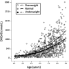
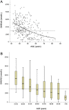
Results: We confirmed that overweight girls exhibited higher and earlier DHEAS levels and no difference was found compared with the average European and South American girls with a similar body mass index (BMI). Adrenopause onset threshold (AOT) defined as DHEAS blood levels <2040 nmol/L was identified in >35% of the females >40 years old and associated with significantly reduced ZR cell number (based on PTEN and hematoxylin signals). ZR cell loss may in part account for lower DHEA/DHEAS expression, but most cells remain alive with lower DHEA/DHEAS biosynthesis.
Conclusion: The timely relation between significant reduction of blood DHEAS levels and decreased ZR cell number at the beginning of the 40s suggests that adrenopause is an additional burden for a significant number of middle-aged women, and may become an emergent problem associated with further sex steroids reduction during the menopausal transition.
Keywords: DHEA-S; DHEAS; PTEN; adrenarche; adrenopause; dehydroepiandrosterone; female; phosphatase and tensin homolog; sulfated-dehydroepiandrosterone.
Figures


Similar articles
-
Adrenopause or decline of serum adrenal androgens with age in women living at sea level or at high altitude.J Endocrinol. 2002 Apr;173(1):95-101. doi: 10.1677/joe.0.1730095. J Endocrinol. 2002. PMID: 11927388
-
The zona reticularis is the site of biosynthesis of dehydroepiandrosterone and dehydroepiandrosterone sulfate in the adult human adrenal cortex resulting from its low expression of 3 beta-hydroxysteroid dehydrogenase.J Clin Endocrinol Metab. 1996 Oct;81(10):3558-65. doi: 10.1210/jcem.81.10.8855801. J Clin Endocrinol Metab. 1996. PMID: 8855801
-
Adrenarche is associated with decreased 3 beta-hydroxysteroid dehydrogenase expression in the adrenal reticularis.Endocr Res. 1996 Nov;22(4):723-8. doi: 10.1080/07435809609043768. Endocr Res. 1996. PMID: 8969933
-
The rise in adrenal androgen biosynthesis: adrenarche.Semin Reprod Med. 2004 Nov;22(4):337-47. doi: 10.1055/s-2004-861550. Semin Reprod Med. 2004. PMID: 15635501 Review.
-
The Enigma of the Adrenarche: Identifying the Early Life Mechanisms and Possible Role in Postnatal Brain Development.Int J Mol Sci. 2021 Apr 21;22(9):4296. doi: 10.3390/ijms22094296. Int J Mol Sci. 2021. PMID: 33919014 Free PMC article. Review.
Cited by
-
Age-related Changes in the Adrenal Cortex: Insights and Implications.J Endocr Soc. 2023 Jul 20;7(9):bvad097. doi: 10.1210/jendso/bvad097. eCollection 2023 Aug 1. J Endocr Soc. 2023. PMID: 37564884 Free PMC article. Review.
-
Human-like adrenal features in Chinese tree shrews revealed by multi-omics analysis of adrenal cell populations and steroid synthesis.Zool Res. 2024 May 18;45(3):617-632. doi: 10.24272/j.issn.2095-8137.2023.280. Zool Res. 2024. PMID: 38766745 Free PMC article.
-
Normal and Premature Adrenarche.Endocr Rev. 2021 Nov 16;42(6):783-814. doi: 10.1210/endrev/bnab009. Endocr Rev. 2021. PMID: 33788946 Free PMC article. Review.
References
-
- Belgorosky A, Baquedano MS, Guercio G, Rivarola MA.Expression of the IGF and the aromatase/estrogen receptor systems in human adrenal tissues from early infancy to late puberty: implications for the development of adrenarche. Reviews in Endocrine and Metabolic Disorders 2009. 10 51–61. (10.1007/s11154-008-9105-1) - DOI - PubMed
LinkOut - more resources
Full Text Sources
Research Materials

