. 2021 Jan;28(1):27-37.
doi: 10.1530/ERC-19-0510.

Serine synthesis influences tamoxifen response in ER+ human breast carcinoma

Affiliations

Serine synthesis influences tamoxifen response in ER+ human breast carcinoma

Stephanie Metcalf et al. Endocr Relat Cancer. 2021 Jan.

Abstract

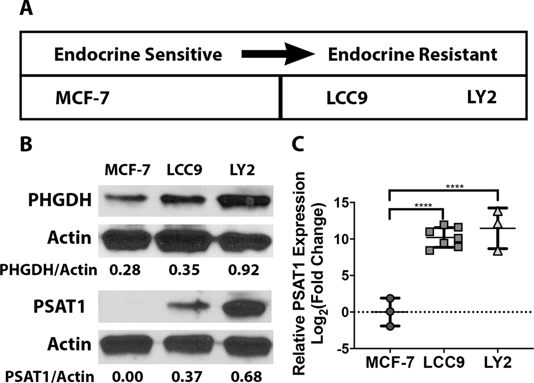
Estrogen receptor-positive breast cancer (ER+ BC) is the most common form of breast carcinoma accounting for approximately 70% of all diagnoses. Although ER-targeted therapies have improved survival outcomes for this BC subtype, a significant proportion of patients will ultimately develop resistance to these clinical interventions, resulting in disease recurrence. Phosphoserine aminotransferase 1 (PSAT1), an enzyme within the serine synthetic pathway (SSP), has been previously implicated in endocrine resistance. Therefore, we determined whether expression of SSP enzymes, PSAT1 or phosphoglycerate dehydrogenase (PHGDH), affects the response of ER+ BC to 4-hydroxytamoxifen (4-OHT) treatment. To investigate a clinical correlation between PSAT1, PHGDH, and endocrine resistance, we examined microarray data from ER+ patients who received tamoxifen as the sole endocrine therapy. We confirmed that higher PSAT1 and PHGDH expression correlates negatively with poorer outcomes in tamoxifen-treated ER+ BC patients. Next, we found that SSP enzyme expression and serine synthesis were elevated in tamoxifen-resistant compared to tamoxifen-sensitive ER+ BC cells in vitro. To determine relevance to endocrine sensitivity, we modified the expression of either PSAT1 or PHGDH in each cell type. Overexpression of PSAT1 in tamoxifen-sensitive MCF-7 cells diminished 4-OHT inhibition on cell proliferation. Conversely, silencing of either PSAT1 or PHGDH resulted in greater sensitivity to 4-OHT treatment in LCC9 tamoxifen-resistant cells. Likewise, the combination of a PHGDH inhibitor with 4-OHT decreased LCC9 cell proliferation. Collectively, these results suggest that overexpression of serine synthetic pathway enzymes contribute to tamoxifen resistance in ER+ BC, which can be targeted as a novel combinatorial treatment option.

Keywords: endocrine resistance; estrogen receptor-positive breast cancer; phosphoserine aminotransferase 1; serine synthesis; tamoxifen.


Conflict of interest statement

Conflict of Interest

All authors declare that there are no conflicts of interest.

Figures

Figure 1:
Figure 1:. Clinical relevance of PSAT1 in Tamoxifen treated ER+BC.
a) Relapse-free survival (RFS) of ER+ patients treated with tamoxifen stratified by above (n=33) or below (n=32) the median PSAT1 expression. Retrospective Kaplan-Meier analysis was performed from microarray data on institutionally collected de-identified laser captured micro-dissected ER+ breast carcinomas from patients treated with tamoxifen. b) Kaplan-Meier analysis of patient outcomes was done using KM Plotter database. RFS of ER+ patients treated with tamoxifen stratified by low (n=73) or high (n=37) PSAT1 expression. Shown are the hazard ratios (HR) and logrank P values for both analyses.
Figure 2:
Figure 2:. Differential PSAT1 expression between sensitive and resistant cell lines.
a) Schematic summarizing the in vitro ER+ breast cancer model systems in terms of responsiveness to endocrine therapy. b) Representative Western blots demonstrating the differential expression of PHGDH and PSAT1 between the endocrine sensitive MCF-7 parental line and the endocrine resistant derivatives (LCC9 and LY2). Densitometry analysis from three independent experiments was performed using ImageJ. c) PSAT1 transcript levels in the endocrine sensitive and resistant cell lines. Quantitation is demonstrated as log2 fold change compared to MCF-7 cells and shown are the mean ± SD from at least three independent experiments. **** P < 0.0001.
Figure 3:
Figure 3:. Metabolomic differences between endocrine sensitive and endocrine resistant cell types.
a) Schematic demonstrating serine/glycine carbon labeling patterns derived from 13C6-glucose incubation. Metabolism of uniformly labeled glucose results in triply labeled (m+3) 3-phosphoglycerate (3-PG). This labeled glycolytic intermediate is then utilized within the SSP to generate triply labeled serine and two-carbon labeled glycine. b) Schematic demonstrating incorporation of glutamine derived nitrogen into the amino group of intracellular metabolized serine and glycine. Incubation of 13C5,15N2-glutamine results in generation of intracellular nitrogen labeled glutamate, which is then converted to alpha-ketoglutarate by PSAT1 through transamination of amino group to form serine. c) 13C-serine (m+3) and 13C-glycine (m+2) levels were quantified by mass spectrometry analysis from polar metabolite extracts from either MCF-7 or LY2 cells after 13C6-glucose incubation. Data is represented as fractional enrichment and shown are the mean ± SD from three independent experiments. d) Polar metabolite extracts from 13C5,15N2-glutamine labeled MCF-7 or LY2 cells were analyzed by mass spectrometry for nitrogen enrichment into 15N-serine (m+1) or 15N-glycine (m+1). Quantitation is demonstrated as fractional enrichment and shown are mean ± SD from three independent experiments.
Figure 4:
Figure 4:. Overexpression of PSAT1 reduces sensitivity to 4-hydroxytamoxifen.
a) Analysis of PSAT1 transcript levels in the MCF-7 cells stably integrated with either empty vector or vector encoding PSAT1 cDNA. Data is presented as log2 fold change and shown is the mean ± SD from at least three independent experiments. *** p < 0.001 as determine by unpaired t test. b) Representative Western blot demonstrating PSAT1 protein levels in MCF-7 cells stably selected for PSAT1 overexpression. Densitometry was performed with Image J from three separate experiments. c) Effect of transient PSAT1 overexpression on LCC9 cell proliferation. Data represented as quantitated cell number (x104) and shown is mean ± SD from four separate experiments. d) Response to 4-OHT of MCF-7 cells with or without overexpression of PSAT1. Data is shown as mean ± SD (from four independent experiments) and represented as % decrease relative to EtOH control. *** P < 0.001 100nM 4-OHT, **** P < 0.0001 500nM 4-OHT.
Figure 5:
Figure 5:. Suppression of PSAT1 increases sensitivity to 4-hydroxytamoxifen.
a) Representative Western blot demonstrating PSAT1 expression in LCC9 cells harboring either scrambled (control) or PSAT1 specific shRNA. Densitometry analysis was performed with Image J from three independent experiments. b) Assessment of PSAT1 loss on LCC9 cell proliferation. Data is presented as total cell number (x104) and shown is the mean ± SD from five independent experiments. *** p < 0.05 as determine by unpaired t test. c) Effect of 4-OHT treatment on cell proliferation in control or PSAT1 silenced LCC9 cells. Quantitation is demonstrated as % decrease relative to EtOH control. Shown is the mean ± SD from five independent experiments. * P < 0.05 250nM 4-OHT, **** P < 0.0001 500nM 4-OHT.
Figure 6:
Figure 6:. Clinical relevance of PHGDH in ER+ patients treated with tamoxifen.
Kaplan-Meier analysis of patient outcomes was done using the online KM Plotter database. Relapse-free survival of ER+ patients treated with tamoxifen stratified by low (n=486) or high (n=184) PHGDH expression. Shown are the hazard ratios (HR) and logrank P values.
Figure 7:
Figure 7:. Suppression of PHGDH increases sensitivity to 4-hydroxytamoxifen.
a) Representative Western blot demonstrating PHGDH expression in LCC9 cells transiently transfected with either scrambled (control) or PHGDH-specific siRNA. Densitometry analysis was performed with Image J. b) Effect of transient PHGDH suppression on cell proliferation in LCC9 cells. Data represented as quantitated cell number (x104) and shown is mean ± SD from three separate experiments. c) Effect of 4-OHT treatment on cell proliferation in control or PSAT1 silenced LCC9 cells. Quantitation is demonstrated as % decrease relative to EtOH control from three independent experiments. Shown is the mean ± SD. ** P < 0.005. d) Effect of PHGDH inhibitor on sensitivity of LCC9 cells to 4-OHT. Data are presented as the mean ± SEM of cell viability relative to DMSO-treated control of four independent experiments. *P < 0.007 versus PKUMDL-WQ-2201 as determined by one-way ANOVA followed by Tukey’s multiple comparisons post hoc test.

Similar articles

Cited by

References

    1. Ali S, Rasool M, Chaoudhry HP,NP, Jha P, Hafiz A, Mahfooz M, Abdus Sami G, Azhar Kamal M, Bashir S, et al. 2016. Molecular mechanisms and mode of tamoxifen resistance in breast cancer. Bioinformation, 12, 135–139. - PMC - PubMed
    1. Andres SA, Brock GN & Wittliff JL 2013. Interrogating differences in expression of targeted gene sets to predict breast cancer outcome. BMC Cancer, 13, 326. - PMC - PubMed
    1. Andres SA & Wittliff JL 2012. Co-expression of genes with estrogen receptor-alpha and progesterone receptor in human breast carcinoma tissue. Horm Mol Biol Clin Investig, 12, 377–90. - PubMed
    1. Astruc ME, Chabret C, Bali P, Gagne D & Pons M 1995. Prolonged treatment of breast cancer cells with antiestrogens increases the activating protein-1-mediated response: involvement of the estrogen receptor. Endocrinology, 136, 824–32. - PubMed
    1. Bai Z & Gust R 2009. Breast cancer, estrogen receptor and ligands. Arch Pharm (Weinheim), 342, 133–49. - PubMed

Publication types