. 2020 Dec;25(8):1129-1138.
doi: 10.1007/s00775-020-01826-8. Epub 2020 Oct 28.

Unexpected photosensitivity of the well-characterized heme enzyme chlorite dismutase

Affiliations

Unexpected photosensitivity of the well-characterized heme enzyme chlorite dismutase

Durga Mahor et al. J Biol Inorg Chem. 2020 Dec.

Abstract

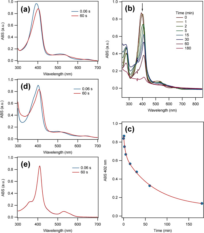
Chlorite dismutase is a heme enzyme that catalyzes the conversion of the toxic compound ClO2- (chlorite) to innocuous Cl- and O2. The reaction is a very rare case of enzymatic O-O bond formation, which has sparked the interest to elucidate the reaction mechanism using pre-steady-state kinetics. During stopped-flow experiments, spectroscopic and structural changes of the enzyme were observed in the absence of a substrate in the time range from milliseconds to minutes. These effects are a consequence of illumination with UV-visible light during the stopped-flow experiment. The changes in the UV-visible spectrum in the initial 200 s of the reaction indicate a possible involvement of a ferric superoxide/ferrous oxo or ferric hydroxide intermediate during the photochemical inactivation. Observed EPR spectral changes after 30 min reaction time indicate the loss of the heme and release of iron during the process. During prolonged illumination, the oligomeric state of the enzyme changes from homo-pentameric to monomeric with subsequent protein precipitation. Understanding the effects of UV-visible light illumination induced changes of chlorite dismutase will help us to understand the nature and mechanism of photosensitivity of heme enzymes in general. Furthermore, previously reported stopped-flow data of chlorite dismutase and potentially other heme enzymes will need to be re-evaluated in the context of the photosensitivity. Illumination of recombinantly expressed Azospira oryzae Chlorite dismutase (AoCld) with a high-intensity light source, common in stopped-flow equipment, results in disruption of the bond between FeIII and the axial histidine. This leads to the enzyme losing its heme cofactor and changing its oligomeric state as shown by spectroscopic changes and loss of activity.

Keywords: Azospira oryzae; Chlorite dismutase; Electron paramagnetic resonance; Heme enzyme; Oligomeric state; Photosensitivity; Stopped-flow spectroscopy; UV–visible illumination.


Conflict of interest statement

There are no conflicts of interest to declare.

Figures

Fig. 1
Fig. 1
The effect of illumination of the stopped-flow (SF) light source on the UV–visible spectra of heme proteins. a SF spectroscopy of 18 µM AoCld in 100 mM KPi pH 7.0 against Milli-Q water in 1:1 ratio at 20 °C. The spectra at 0.06 s (blue) and 60 s (red) are given. b UV–visible spectroscopy of 16 µM AoCld in 100 mM KPi pH 7.0 mixed 1:1 with Milli-Q water at 20 °C after different illumination times by the Xenon arc lamp. c Decay of the Soret peak at 402 nm of AoCld during the illumination experiment under (b). The solid red line is a fit to a double exponential decay with a first-order rate constant kdecay,1 = (0.43 ± 0.35)·10–2 s−1 and kdecay,2 = (2.3 ± 1.2)·10–4 s−1 (details of the fit are given in the supplementary information). d SF spectroscopy of 20 µM Horseradish peroxidase in water against Milli-Q water in 1:1 ratio at 20 °C. The spectra at 0.06 s (blue) and 60 s (red) are given. e SF spectroscopy of 17 µM Bovine heart Cytochrome c (oxidized) in water against Milli-Q water in 1:1 ratio at 20 °C. The spectra at 0.06 s (blue) and 60 s (red) are given
Fig. 2
Fig. 2
Kinetic analysis of SF spectroscopy of AoCld versus 1 equivalent (eq.) chlorite using singular value decomposition (SVD) assuming an irreversible first-order reaction. a Reconstructed spectra obtained after SVD analysis and fitting. In blue is the ferric resting state and in red is the photoinactived species. b Reconstructed spectra focussed on Q-bands. In blue is the ferric resting state and in red is the photoinactived species. c Kinetic traces in grey represent the reconstructed traces from the SVD analysis and the red and blue traces represent the fit to an irreversible first-order reaction with k1 = (1.51 ± 0.05)·10–2 s−1. SF experimental conditions: 18 µM AoCld in 100 mM KPi pH 7.0 against 18 μM sodium chlorite (1 eq.) in water in 1:1 ratio at 20 °C
Fig. 3
Fig. 3
Characterization of AoCld oligomeric state, activity and EPR properties after illumination. a The specific activity of non-illuminated (0 min), 30 min, 60 min and 180 min illuminated 135 pM AoCld in 100 mM KPi pH 7.0 and 1 mM sodium chlorite at 21 °C. The red line is a double exponential curve with rate constants as determined for the Soret decay, with only the two amplitudes of the two phases as fitparameters. 100% activity represents a specific activity of (8.53 ± 0.17)·103 U/mg b EPR spectra of non-illuminated and illuminated AoCld: 75 µM of non-illuminated (blue) and the same concentration of protein illuminated for 30 min (green), 60 min (brown) and 180 min (purple) prior to freezing. EPR conditions: microwave frequency, 9.405 GHz, microwave power, 20 mW, modulation frequency, 100 kHz; modulation amplitude, 10 Gauss; temperature, 20 K. c Analytical size exclusion chromatography was performed before illumination (0 min) and after 30 min, 60 min and 180 min illumination. All gel chromatograms were recorded at two different wavelengths 280 nm (blue) and 410 nm (red). Calibration of the column is given in supplemental Fig. S4

References

    1. Hofbauer S, Schaffner I, Furtmüller PG, Obinger C. Biotechnol J. 2014;9:461–473. doi: 10.1002/biot.201300210. - DOI - PMC - PubMed
    1. Mlynek G, Sjöblom B, Kostan J, Füreder S, Maixner F, Gysel K, Furtmüller PG, Obinger C, Wagner M, Daims H, Djinović-Carugo K. J Bacteriol. 2011;193:2408–2417. doi: 10.1128/JB.01262-10. - DOI - PMC - PubMed
    1. Mobilia KC, Hutchison JM, Zilles JL. Front Microbiol. 2017 doi: 10.3389/fmicb.2017.02423. - DOI - PMC - PubMed
    1. Lee AQ, Streit BR, Zdilla MJ, Abu-Omar MM, DuBois JL. Proc Natl Acad Sci. 2008;105:15654–15659. doi: 10.1073/pnas.0804279105. - DOI - PMC - PubMed
    1. Mehboob F, Wolterink AFM, Vermeulen AJ, Jiang B, Hagedoorn P-L, Stams AJM, Kengen SWM. FEMS Microbiol Lett. 2009;293:115–121. doi: 10.1111/j.1574-6968.2009.01517.x. - DOI - PubMed

Publication types

Supplementary concepts

LinkOut - more resources