. 2020 Dec;52(12):4824-4839.
doi: 10.1111/ejn.15027. Epub 2020 Nov 11.

Comparative analyses of transgene expression patterns after intra-striatal injections of rAAV2-retro in rats and rhesus monkeys: A light and electron microscopic study

Affiliations

Comparative analyses of transgene expression patterns after intra-striatal injections of rAAV2-retro in rats and rhesus monkeys: A light and electron microscopic study

Daniel L Albaugh et al. Eur J Neurosci. 2020 Dec.

Abstract

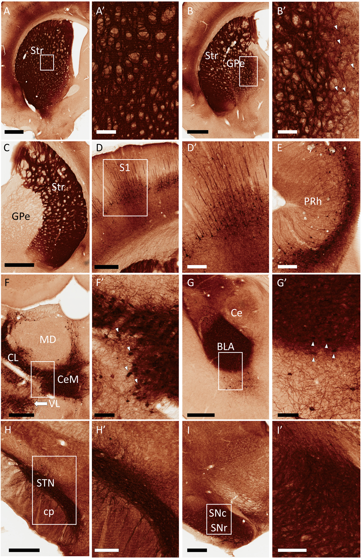
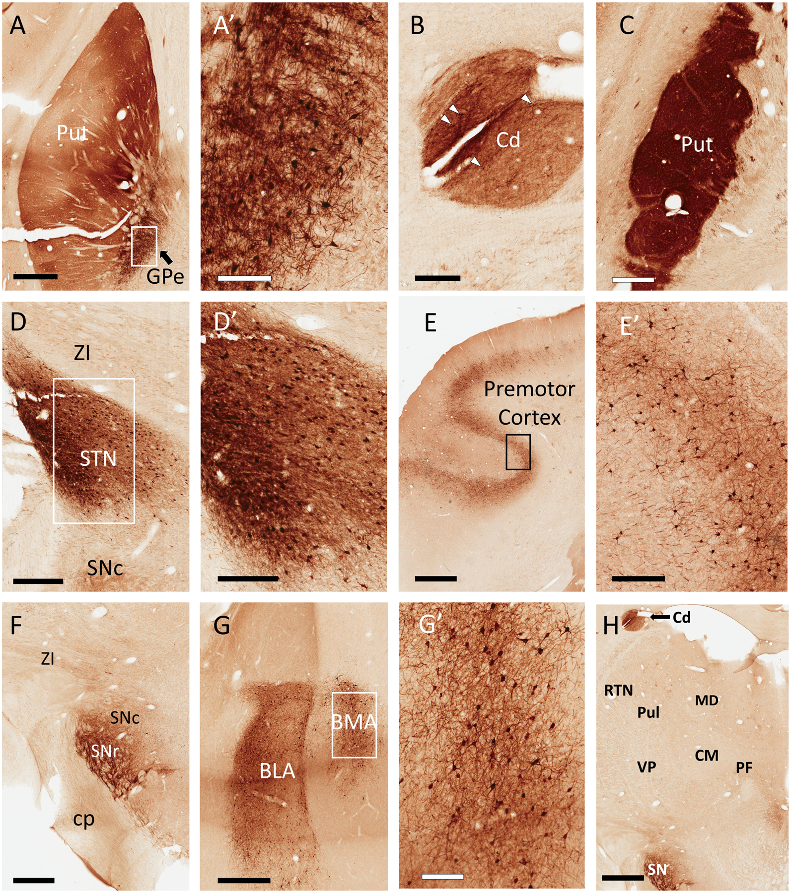
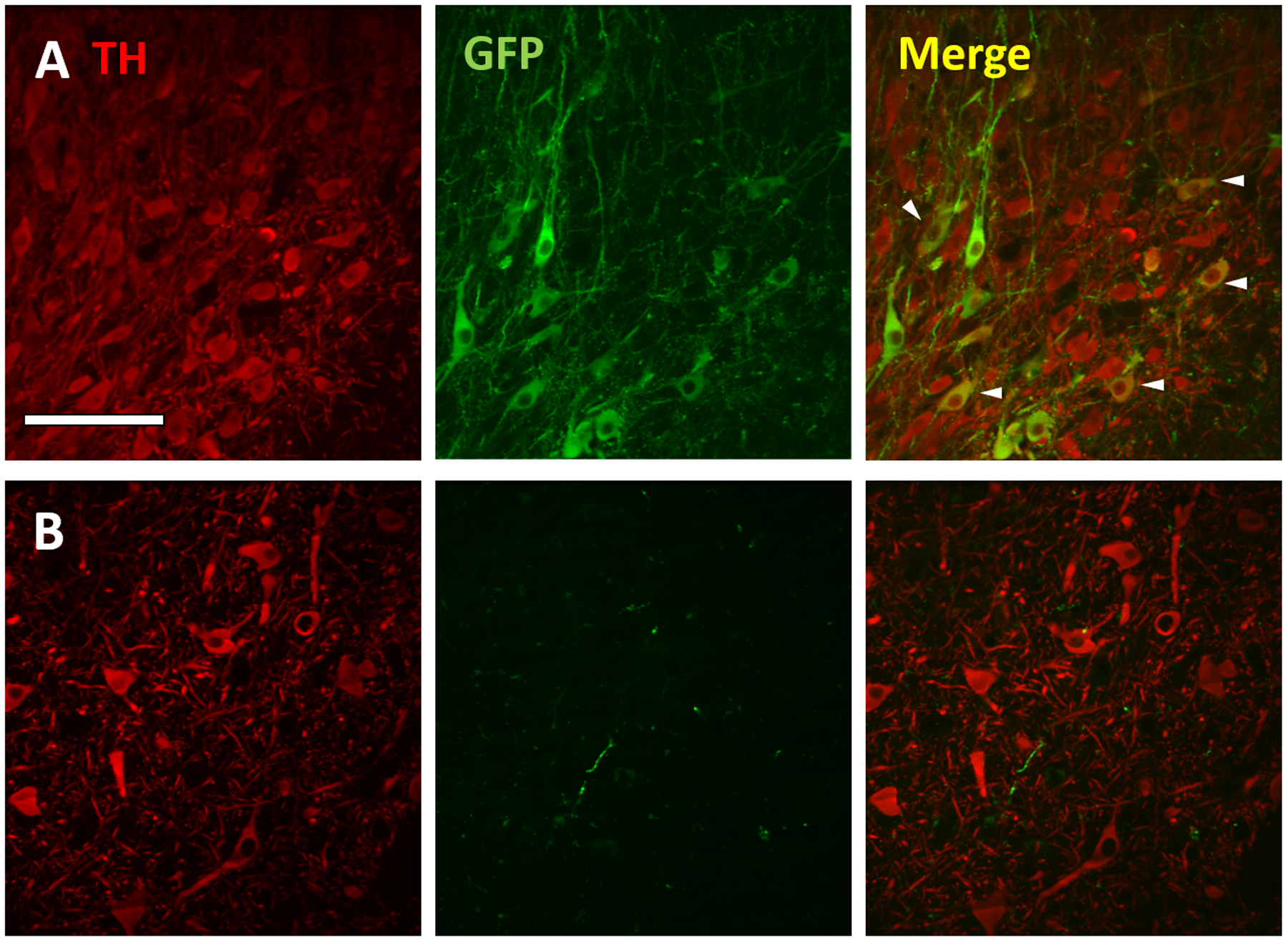
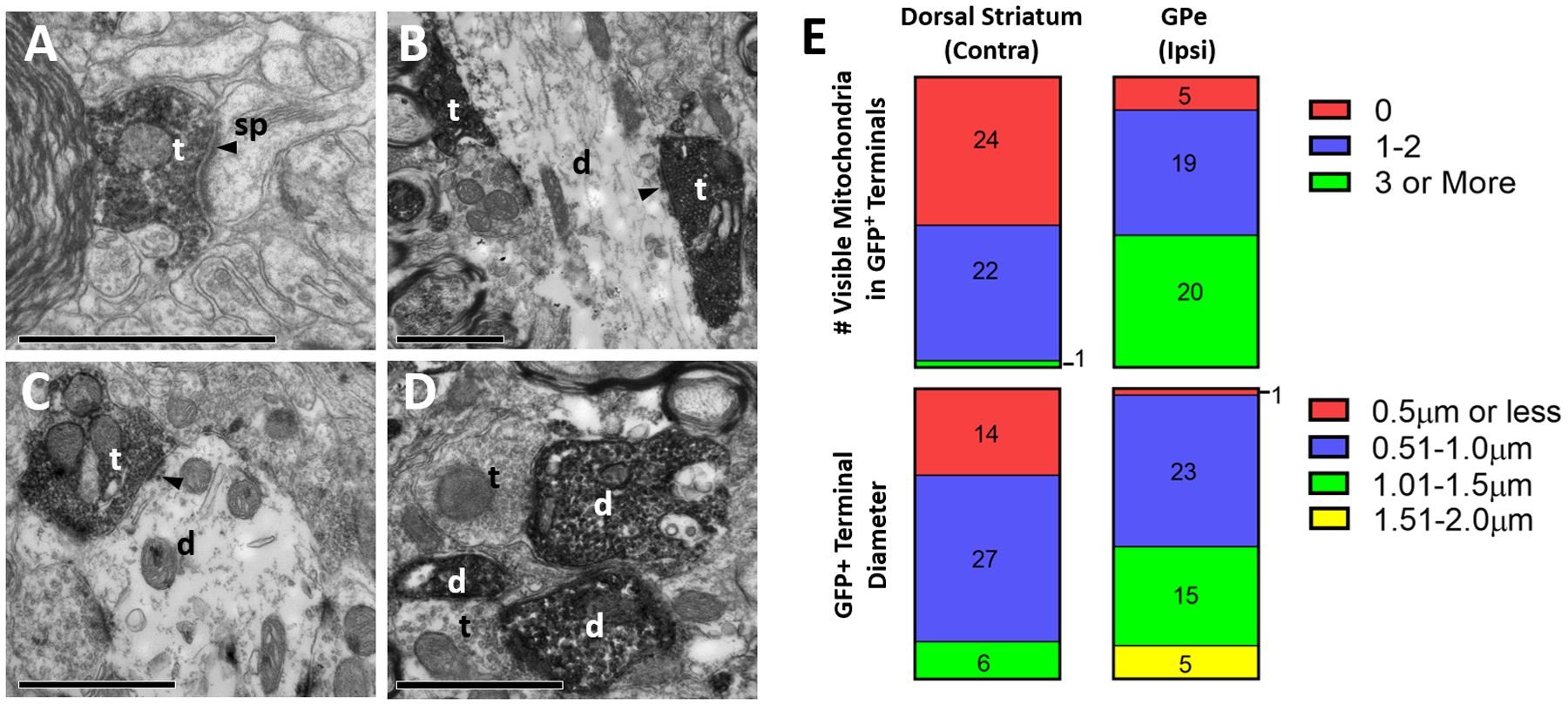
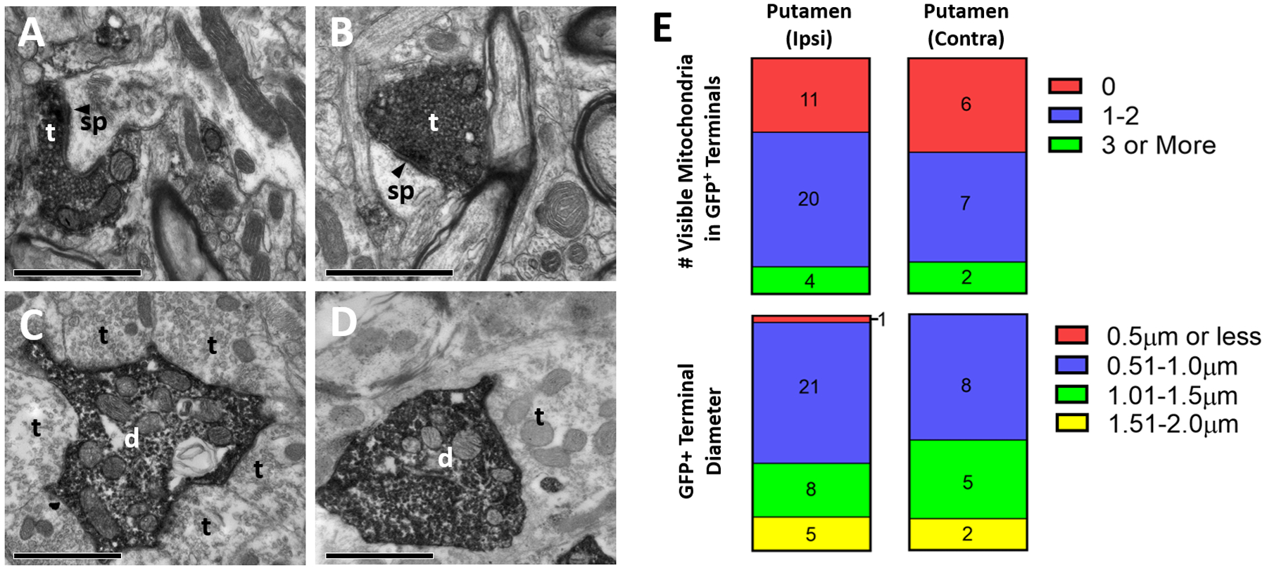
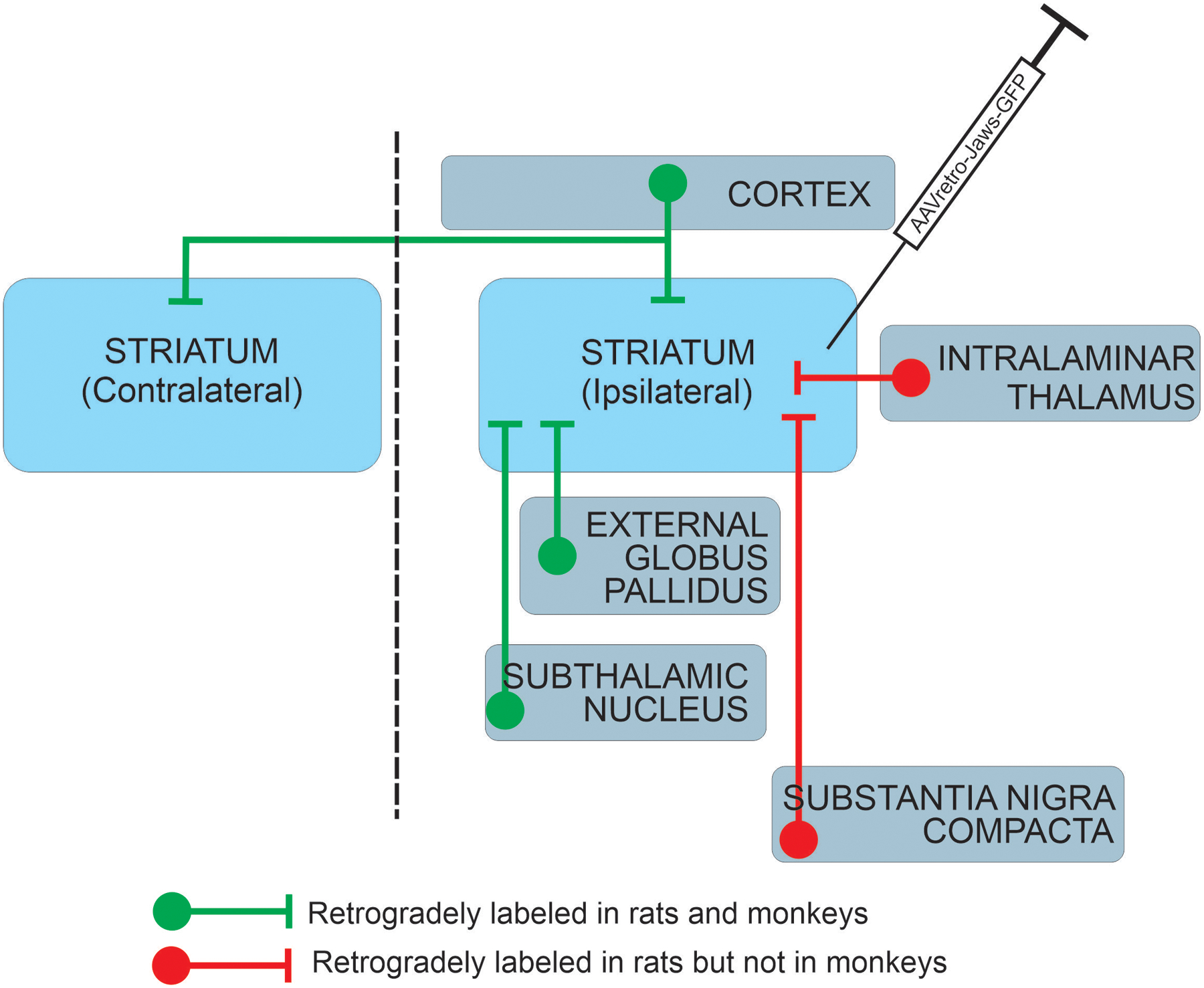
Retrogradely-transducing viral vectors are versatile tools for anatomical and functional interrogations of neural circuits. These vectors can be applied in nonhuman primates (NHPs), powerful model species for neuroscientific studies with limited genetic tractability, but limited data are available regarding the tropism and transgene expression patterns of such viruses after injections in NHP brains. Consequently, NHP researchers must often rely on related data available from other species for experimental planning. To evaluate the suitability of rAAV2-retro in the NHP basal ganglia, we studied the transgene expression patterns at the light and electron microscope level after injections of rAAV2-retro vector encoding the opsin Jaws conjugated to a green fluorescent protein (GFP) in the putamen of rhesus macaques. For inter-species comparison, we injected the same vector in the rat dorsal striatum. In both species, GFP expression was observed in numerous cortical and subcortical regions with known striatal projections. However, important inter-species differences in pathway transduction were seen, including labeling of the intralaminar thalamostriatal projection in rats, but not monkeys. Electron microscopic ultrastructural observations within the basal ganglia revealed GFP labeling in both postsynaptic dendrites and presynaptic axonal terminals; the latter likely derived from anterograde transgene transport in neurons that project to the striatum, and from collaterals of these neurons. Our results suggest that certain neural pathways may be refractory to transduction by retrograde vectors in a species-specific manner, highlighting the need for caution when determining the suitability of a retrograde vector for NHP studies based solely on rodent data.

Keywords: connectome; corticostriatal; electron microscopy; primate; retrograde transduction; striatum; subthalamic nucleus; thalamostriatal; viral vectors.


Figures

Figure 1:
Figure 1:
Representative light micrographs of GFP expression in rat brain following a unilateral, intra-striatal injection of rAAV2-retro-hSyn-Jaws-GFP. All regions shown are from the hemisphere ipsilateral to the virus injection, except where indicated. Areas in rectangles are shown at higher magnification in the corresponding panels labeled with prime symbols. (A) Striatum (Str), (B) External globus pallidus (GPe), (C) GPe, contralateral side, (D) Ipsilateral primary somatosensory cortex (S1), (E) Perirhinal cortex (PRh), (F) Thalamus, (G) Amygdala, (H) Subthalamic nucleus (STN), (I) Substantia nigra. Additional abbreviations: basolateral amygdala (BLA), central amygdala (Ce), centrolateral thalamus (CL), central medial thalamus (CeM), cerebral peduncle (cp), mediodorsal thalamus (MD), substantia nigra pars reticulata (SNr), substantia nigra pars compacta (SNc), ventrolateral thalamus (VL). Arrowheads in B’, F’ and G’ point to subsets of GFP+ cell bodies visible in these panels. GFP+ cells were also numerous in the STN, and also present in the SNc, although the intensity of neuropil labeling in these regions rendered such cell bodies difficult to display by these peroxidase images (see Figs. 3 and 4 for evidence of cell labeling in the rat SNc and STN, respectively). Scale Bars: 1mm (A, B, C); 500μm (D, G, H, I); 300μm (F); 200μm (A’, B’, D’, E, H’, I’); 100μm (F’, G’). Approximate rostrocaudal coordinates of sections, according to (Paxinos & Watson, 2006) in mm relative to Bregma: 1.2 (A); −0.84 (B, C, D); −4.44 (E); −2.52 (F); −3.36 (G); −3.6 (H); −6.12 (I). Figure includes micrographs from all 3 rats used in this study.
Figure 2:
Figure 2:
Representative light micrographs of GFP expression in monkey brain following a unilateral, intra-striatal (putamen) injection of rAAV2-retro-hSyn-Jaws-GFP. All regions described below were ipsilateral to injection site. In the putamen (Put), neuropil was diffusely labeled, with scattered GFP+ cell bodies, particularly in the ventral territory (A). In the same section, numerous GFP+ cell bodies were found in the external globus pallidus (GPe) (A and A’). Similar to the Put, GFP+ fiber staining was observed in the caudate (Cd) (B; note that this section of caudate is also visible in Panel H). Similar to the rat striatum, the neuropil of the Put was densely labeled across the rostrocaudal axis, many millimeters away from the injection site (C). The subthalamic nucleus (STN) contained numerous GFP+ cell bodies and dense neuropil staining (D and D’). Varying numbers of deep-layer pyramidal cells were labeled in many cortical regions, including the premotor cortex (E, E’). In the substantia nigra, we observed GFP+ fibers, as well as a few labeled cell bodies bordering the compacta (SNc) and reticulata (SNr) territories (F). Numerous GFP+ cells were located in the basolateral and basomedial amygdala (BLA and BMA, respectively) (G and G’). Remarkably, the thalamus was nearly devoid of GFP labeling (H). Additional abbreviations: centromedian nucleus of thalamus (CM), parafascicular nucleus of thalamus (PF), pulvinar (Pul), thalamic reticular nucleus (RTN), ventral posterior nucleus of thalamus (VP), zona incerta (ZI), all other abbreviations as described in Figure 1. Scale Bars: 2mm (E, H) 1mm (A, F, G); 600μm (C); 500μm (D); 300μm (B, D’, E’); 200μm (A’, G’). Approximate rostrocaudal coordinates of sections, according to (Paxinos et al., 1999) in mm relative to interaural line: 10.2 (A, D); 7.5 (B, C, F, H); 19.65 (E); 14.7 (G). Figure includes micrographs from both monkeys used in this study.
Figure 3:
Figure 3:
GFP expression in dopaminergic (tyrosine hydroxylase (TH)-positive) neurons in the rat, but not monkey SNc following intra-striatal rAAV2-retro injection. Confocal images of TH (red; left), GFP (green; middle) and merged markers (right) of the rat (row A) and monkey (row B). SNc ipsilateral to injection site. Arrowheads in merged image depict a subset of GFP+/TH+ cells (yellow) in the rat SNc. Scale bar: 100μm.
Figure 4:
Figure 4:
Ultrastructural analysis of GFP-positive elements in the rat basal ganglia, after intra-striatal injection of rAAV2-retro-hSyn-Jaws-GFP. A-D: Representative electron micrographs of GFP-immunopositive elements (revealed with peroxidase). (A) GFP-labeled axonal terminal in striatum contralateral to the injection site, (B) labeled terminals in GPe ipsilateral to the injection site, (C-D) labeled terminal and dendrites in ipsilateral STN. Abbreviations: t (axon terminal), d (dendrite), sp (spine). White or black text for these abbreviations indicate if the element is GFP-positive or negative, respectively. Arrowhead denotes asymmetric synapse made by labeled terminals. All scale bars: 1μm. (E) Quantification of diameter (top) and number of visible mitochondria inside immunolabeled terminals (bottom) in the contralateral striatum (left) and ipsilateral GPe (right). Numbers in bars refer to total terminal counts for each category.
Figure 5:
Figure 5:
Ultrastructural analysis of GFP-positive elements in the monkey basal ganglia after intra-striatal injection of rAAV2-retro-hSyn-Jaws-GFP. A-D: Representative electron micrographs of GFP-immunopositive elements (revealed with peroxidase). (A) GFP-positive terminal in putamen ipsilateral to injection site, (B) labeled terminal in putamen contralateral to injection site, (C) labeled dendrite in ipsilateral GPe, (D) labeled dendrite in ipsilateral STN. Abbreviations: t (axon terminal), d (dendrite), sp (spine). White or black text for these abbreviations indicate if the element is GFP-positive or negative, respectively. Arrowhead denotes asymmetric synapse made by labeled terminals. All scale bars: 1μm. (E) Quantification of diameter (top) and number of visible mitochondria inside immunolabeled terminals (bottom) in the ipsilateral (left) and contralateral (right) putamen. Numbers in bars refer to total terminal counts for each category.
Figure 6:
Figure 6:
Comparison of neuronal pathways transduced by intra-striatal rAAV2-retro injection in rat and monkey.

References

    1. Albaugh DL, Huang C, Ye S, Paré JF & Smith Y (2020) Glutamatergic inputs to GABAergic interneurons in the motor thalamus of control and parkinsonian monkeys. Eur. J. Neurosci - PubMed
    1. Albert K, Voutilainen MH, Domanskyi A, Piepponen TP, Ahola S, Tuominen RK, Richie C, Harvey BK & Airavaara M (2019) Downregulation of tyrosine hydroxylase phenotype after AAV injection above substantia nigra: Caution in experimental models of Parkinson’s disease. J. Neurosci. Res, 97, 346–361. - PMC - PubMed
    1. Berendse HW & Groenewegen HJ (1990) Organization of the thalamostriatal projections in the rat, with special emphasis on the ventral striatum. J. Comp. Neurol, 299, 187–228. - PubMed
    1. Chatterjee S, Sullivan HA, MacLennan BJ, Xu R, Hou Y, Lavin TK, Lea NE, Michalski JE, Babcock KR, Dietrich S, Matthews GA, Beyeler A, Calhoon GG, Glober G, Whitesell JD, Yao S, Cetin A, Harris JA, Zeng H, Tye KM, Reid RC & Wickersham IR (2018) Nontoxic, double-deletion-mutant rabies viral vectors for retrograde targeting of projection neurons. Nat. Neurosci, 21, 638–646. - PMC - PubMed
    1. Chuong AS, Miri ML, Busskamp V, Matthews GA, Acker LC, Sorensen AT, Young A, Klapoetke NC, Henninger MA, Kodandaramaiah SB, Ogawa M, Ramanlal SB, Bandler RC, Allen BD, Forest CR, Chow BY, Han X, Lin Y, Tye KM, Roska B, Cardin JA & Boyden ES (2014) Noninvasive optical inhibition with a red-shifted microbial rhodopsin. Nat. Neurosci, 17, 1123–1129. - PMC - PubMed

Publication types