. 2020 Dec 15;142(24):2356-2370.
doi: 10.1161/CIRCULATIONAHA.120.047239. Epub 2020 Oct 28.

Epigenetic Reader BRD4 (Bromodomain-Containing Protein 4) Governs Nucleus-Encoded Mitochondrial Transcriptome to Regulate Cardiac Function

Affiliations

Epigenetic Reader BRD4 (Bromodomain-Containing Protein 4) Governs Nucleus-Encoded Mitochondrial Transcriptome to Regulate Cardiac Function

Soo Young Kim et al. Circulation. .

Abstract

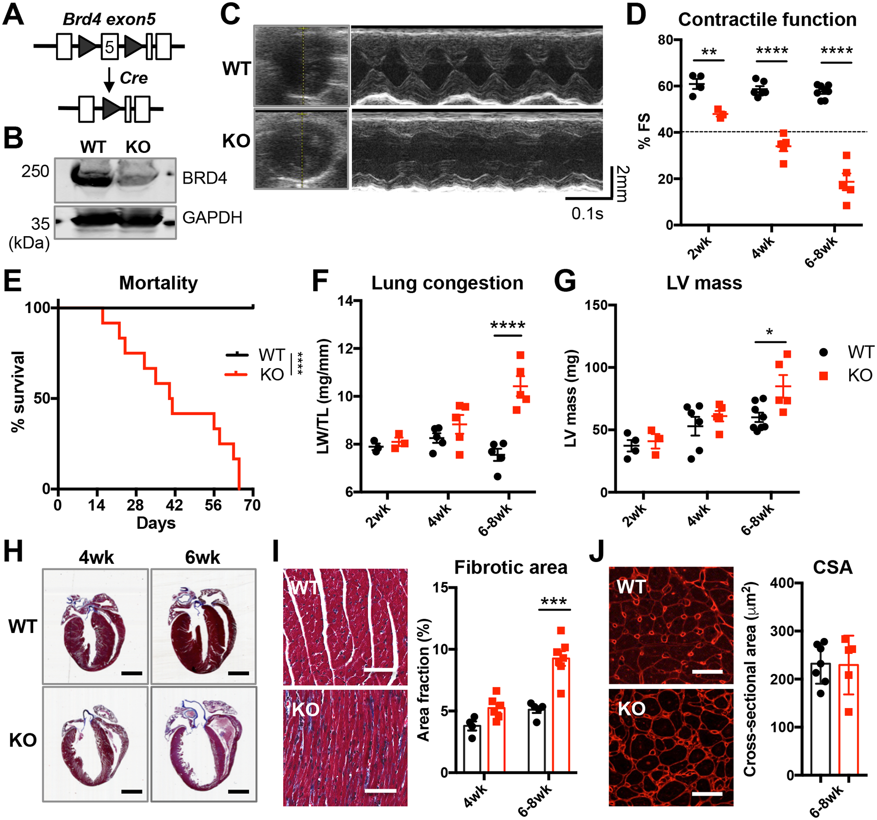
Background: BET (bromodomain and extraterminal) epigenetic reader proteins, in particular BRD4 (bromodomain-containing protein 4), have emerged as potential therapeutic targets in a number of pathological conditions, including cancer and cardiovascular disease. Small-molecule BET protein inhibitors such as JQ1 have demonstrated efficacy in reversing cardiac hypertrophy and heart failure in preclinical models. Yet, genetic studies elucidating the biology of BET proteins in the heart have not been conducted to validate pharmacological findings and to unveil potential pharmacological side effects.

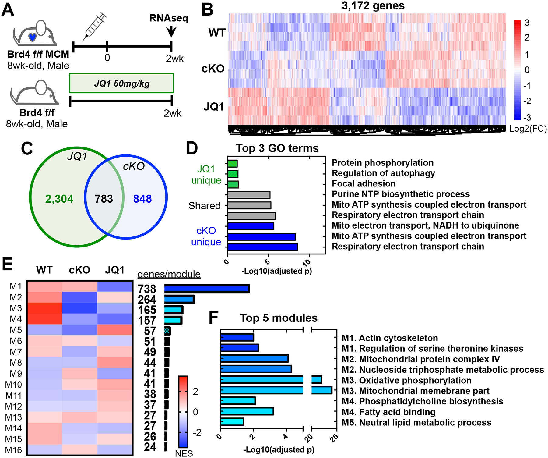
Methods: By engineering a cardiomyocyte-specific BRD4 knockout mouse, we investigated the role of BRD4 in cardiac pathophysiology. We performed functional, transcriptomic, and mitochondrial analyses to evaluate BRD4 function in developing and mature hearts.

Results: Unlike pharmacological inhibition, loss of BRD4 protein triggered progressive declines in myocardial function, culminating in dilated cardiomyopathy. Transcriptome analysis of BRD4 knockout mouse heart tissue identified early and specific disruption of genes essential to mitochondrial energy production and homeostasis. Functional analysis of isolated mitochondria from these hearts confirmed that BRD4 ablation triggered significant changes in mitochondrial electron transport chain protein expression and activity. Computational analysis identified candidate transcription factors participating in the BRD4-regulated transcriptome. In particular, estrogen-related receptor α, a key nuclear receptor in metabolic gene regulation, was enriched in promoters of BRD4-regulated mitochondrial genes.

Conclusions: In aggregate, we describe a previously unrecognized role for BRD4 in regulating cardiomyocyte mitochondrial homeostasis, observing that its function is indispensable to the maintenance of normal cardiac function.

Keywords: BRD4 protein, human; electron transport; epigenetics; heart failure; mitochondria; transcription, genetic.


Conflict of interest statement

DISCLOSURES

The authors declare no competing financial interests.

Figures

Figure 1.
Figure 1.. Cardiomyocyte-specific silencing of BRD4 in developing heart results in post-natal dilated cardiomyopathy.
(A) Schematic of BRD4 conditional allele. (B) Representative immunoblot of BRD4 in WT versus KO LV lysates at 4wks age. (C) Representative echo image at 4wks age, B-mode and M-mode. (D) Echocardiographic analysis of WT and BRD4 cKO at 2wks, 4wks, and 6–8wks of age (n=3–5/group). **p<0.01, ***p<0.005, ****p<0.001 versus age-matched WT. (E) Survival curve of BRD4 CM-deletion mouse (n=11–12/group). ****p<0.001 versus WT. (F) Lung congestion measured by wet lung weight normalized to tibia length over at 2wks, 4wks, and 6–8wks of age (n=3–5/group). ****p<0.001 versus WT. (G) Left ventricular (LV) mass measured by echo at 2wks, 4wks, and 6–8wks (n=3–5/group). *p<0.05 versus WT. (H) Representative 4-chamber cross-section at 4wks and 6wks of age, trichrome stained. Scale bar=20mm. (I, J) Representative images and quantitation of cardiac fibrosis by trichrome staining at 4wks to 8wks of age (n=5–8/group), scale bar=100μm, ***p<0.005 versus WT, and cardiac fiber size by WGA staining at 6–8wks of age (n=5–8/group), scale bar=25μm.
Figure 2.
Figure 2.. Cardiomyocyte-specific deletion of BRD4 in adult heart results in heart failure.
(A) Representative immunoblot for BRD4 in WT versus KO LV lysates at 2wks post tamoxifen injection (arrow indicates the BRD4-FL band). (B) Representative echo image at 5wks post-Tam, B-mode and M-mode. (C) Echocardiographic analysis of WT and BRD4 cKO at 2wks, 3wks, 5wks, and 7wks post-Tam (n=3–7/group). ****p<0.001 versus WT. (D) Survival curve of BRD4 CM-deletion mouse (n=10–14/group). **p<0.01 versus WT. (E) Lung congestion measured by wet lung weight normalized to tibia length 5wks and 7wks post-Tam (n=6–8/group). ***p<0.005 versus WT. (F) Representative images and quantitation of cardiac fiber size by WGA stain (n=6–8/group), scale bar=25μm. *p<0.05, **p<0.01 versus WT. (G) Representative 2-chamber cross-section, Trichrome-stained at 5wks and 7wks post-Tam, and quantifications of tissue fibrosis (n=6–8/group).
Figure 3.
Figure 3.. BRD4-deletion versus inhibition reveals shared and unique transcriptomic profiles.
(A) Schematic for RNAseq sample collection with three groups: WT, cKO at 2wks post induction, and JQ1 after 2wks of treatment (n=4/group). (B) Heatmap of differentially expressed genes between all groups (adj. p<0.05). (C, D) Venn diagram between cKO vs WT and JQ1 vs WT groups and top 3 GO terms for each compartment (adj. p<0.05). (E) Heatmap of the module analysis for differentially expressed genes between all groups (FC>1.5, p<0.05) with total number of genes per module. (F) Significant GO terms for top 5 gene modules.
Figure 4.
Figure 4.. Transcriptome analysis of BRD4-null heart reveals early and significant changes in nucleus-encoded mitochondrial gene network involved in the electron transport chain.
(A) Volcano plot of differentially expressed coding and non-coding genes between WT and cKO groups (FC>1.5, adj. p<0.05). (B) Top 3 GO terms, up or down-regulated in cKO versus WT (FC>1.5, adj. p<0.05). (C) Top enriched gene set form GSEA for cKO versus WT against Hallmark gene sets, adj. p<0.05 represents statistical significance. (D) Metabolic network analysis of the altered genes in cKO versus WT (FC>1.5, adj. p<0.05). Nodes labeled by top ontology terms from HMDB. (E) qPCR validation of RNAseq-identified cKO specific down and up-regulated genes in αMHC-Cre lines at 4wks of age and αMHC-MerCreMer at 5wks post-Tam (n=4–5/group); all shown genes are significantly altered in comparison to WT (p<0.05 by Two-way ANOVA).
Figure 5.
Figure 5.. BRD4 deletion in cardiomyocytes leads to mitochondrial electron transport chain dysfunction.
(A) Significant RNA-seq transcript changes of ETC and TCA genes in cKO versus WT. (B) Schematic of mitochondrial ETC and Schematic of animal models for ventricular mitochondria isolation: WT and αMHC cKO at 4wks of age, and Sham and TAC 5wks post-surgery. (C) Contractile function by echo of samples for mitochondrial enzyme activity assays, αMHC cKO and TAC sets (n=5–8/group). *p<0.05, ***p<0.005, ****p<0.001 versus WT or Sham. (D) Maximal NADH oxidase activity measured by NADH consumption rate of isolated ventricular mitochondria at RT (n=5–8/group). ***p<0.005, ****p<0.001 versus WT or BRD4 KO. (E) Individual ETC complex maximal activity measurement of isolated ventricular mitochondria at RT (n=8/group). *p<0.05, **p<0.01, ***p<0.005 versus WT. (F) TCA cycle enzyme maximal activity of isolated mitochondria at RT. (n=8/group). *p<0.05, **p<0.01 versus WT. (G) Immunoblot of mitochondrial proteins and enzymes in isolated ventricular mitochondria from αMHC cKO and TAC sets. (H) Oxygen consumption measurement of isolated mitochondria at 37°C using different substrates, palmitoyl-carnitine and pyruvate (n=5/group). ****p<0.001 versus WT.
Figure 6.
Figure 6.. Summary figure:
Cardiomyocyte BRD4 plays an indispensable role in driving cardiac gene expression, in particular, nucleus-encoded mitochondrial respiratory enzymes. Without BRD4, cardiomyocyte ETC function deteriorates due to depletion of key enzymes, resulting in depressed respiration and ΔΨM (mitochondrial membrane potential). BRD4 cKO hearts progress to severe contractile dysfunction and heart failure.

References

    1. Gillette TG and Hill JA. Readers, writers, and erasers: chromatin as the whiteboard of heart disease. Circ Res. 2015;116:1245–1253. - PMC - PubMed
    1. Kim SY, Morales CR, Gillette TG and Hill JA. Epigenetic regulation in heart failure. Curr Opin Cardiol. 2016;31:255–265. - PMC - PubMed
    1. Backs J and Olson EN. Control of cardiac growth by histone acetylation/deacetylation. Circ Res. 2006;98:15–24. - PubMed
    1. Anand P, Brown JD, Lin CY, Qi J, Zhang R, Artero PC, Alaiti MA, Bullard J, Alazem K, Margulies KB et al. BET bromodomains mediate transcriptional pause release in heart failure. Cell. 2013;154:569–582. - PMC - PubMed
    1. Spiltoir JI, Stratton MS, Cavasin MA, Demos-Davies K, Reid BG, Qi J, Bradner JE and McKinsey TA. BET acetyl-lysine binding proteins control pathological cardiac hypertrophy. J Mol Cell Cardiol. 2013;63:175–179. - PMC - PubMed

Publication types

MeSH terms