. 2020 Oct 27;33(4):108311.
doi: 10.1016/j.celrep.2020.108311.

Sox2 and Canonical Wnt Signaling Interact to Activate a Developmental Checkpoint Coordinating Morphogenesis with Mesoderm Fate Acquisition

Affiliations

Sox2 and Canonical Wnt Signaling Interact to Activate a Developmental Checkpoint Coordinating Morphogenesis with Mesoderm Fate Acquisition

Brian A Kinney et al. Cell Rep. .

Abstract

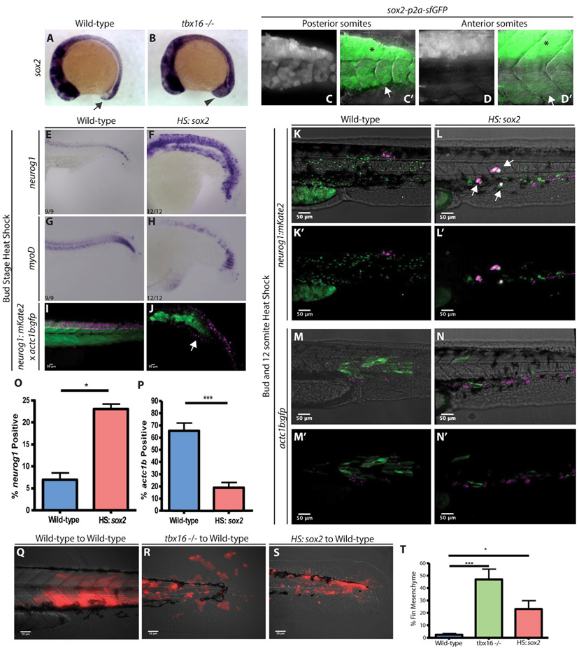
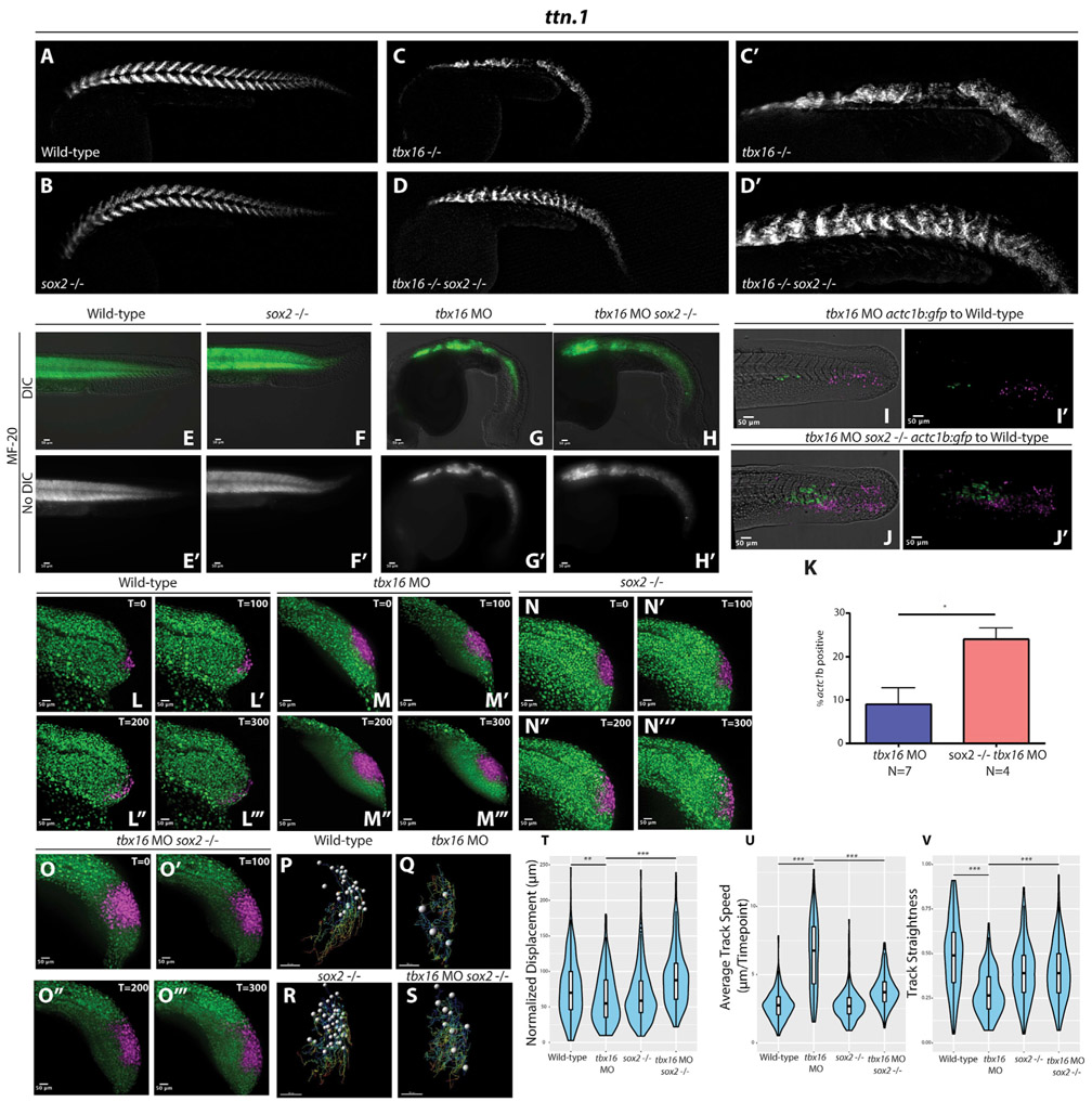
Animal embryogenesis requires a precise coordination between morphogenesis and cell fate specification. During mesoderm induction, mesodermal fate acquisition is tightly coordinated with the morphogenetic process of epithelial-to-mesenchymal transition (EMT). In zebrafish, cells exist transiently in a partial EMT state during mesoderm induction. Here, we show that cells expressing the transcription factor Sox2 are held in the partial EMT state, stopping them from completing the EMT and joining the mesoderm. This is critical for preventing the formation of ectopic neural tissue. The mechanism involves synergy between Sox2 and the mesoderm-inducing canonical Wnt signaling pathway. When Wnt signaling is inhibited in Sox2-expressing cells trapped in the partial EMT, cells exit into the mesodermal territory but form an ectopic spinal cord instead of mesoderm. Our work identifies a critical developmental checkpoint that ensures that morphogenetic movements establishing the mesodermal germ layer are accompanied by robust mesodermal cell fate acquisition.

Keywords: canonical Wnt signaling; mesoderm; neuromesodermal progenitors; somite; sox2; spinal cord; tbx16; zebrafish.


Conflict of interest statement

Declaration of Interests The authors declare no competing interests.

Figures

Figure 1.
Figure 1.. sox2 Activation Causes an Increase of Neural Progenitors and a Decrease in Presomitic Mesoderm
(A and B)12-somite-stage embryos express sox2 in NMPs (A, arrow) and sox2 expression is expanded in tbx16 mutants (B, arrowhead). (C and D) A sox2 reporter line shows perdurance of sfGFP in the most recently formed somites at 24 hpf (C, C’, arrow, spinal cord expression in dorsal region labeled with asterisk), which is absent in more anterior somites (D, D’, arrow, spinal cord expression labeled with asterisk). (E–H) Whole-mount in situ hybridization visualizing neurog1 (neural) (E and F) or myod (skeletal muscle) (G and H) in wild type (E and G) and HS:sox2 embryos (F and H). All of the embryos for in situ hybridization were heat shocked at the bud stage at 40°C for 30 min and fixed at 24 hpf. (I and J) Transgenic embryos with the neurog1:mKate2 and actc1b:gfp reporters show ectopic neural expansion (arrow) and muscle loss in HS:sox2 (J) embryos compared to wild type (I). Live-imaged transgenic embryos were heat shocked at the bud stage at 40°C for 30 min and imaged at 36 hpf. (K–P) Embryos with the neurog1:mkate (K–L’) or the actc1b:gfp (M–N’) reporter were injected with NLS-KikGR mRNA, and cells from these embryos were transplanted into the ventral margin of wild-type host embryos. Donor cells with the HS:sox2 transgene exhibited an increase in the percentage of neurog1:mkate+ cells that also appeared in ectopic locations outside the spinal cord domain (arrows, L, L’, compared to K, K’ and quantified in O; 1,177 wild-type donor cells were counted in 8 host embryos, and 2,051 HS:sox2 donor cells were counted from 10 host embryos; statistics were performed using an unpaired t test, *p = 0.0105) and a decrease in the percentage of actc1b:gfp+ cells (N, N’ compared to M, M’ and quantified in P; 1,307 wild-type donor cells were counted in 8 host embryos, and 971 HS:sox2 donor cells were counted from 5 host embryos; statistics were performed using an unpaired t test, ***p = 0.0003). The NLS-KikGR protein was photoconverted to red fluorescence in (M)–(N’). (Q–T) Wild-type, tbx16 mutant, and HS:sox2 embryos were injected with rhodamine dextran and transplanted into the ventral margin of shield stage wild-type host embryos. The percentage of transplanted cell contribution to fin mesenchyme is quantified in (T) (2,129 wild-type donor cells were counted in 6 host embryos; 2,714 tbx6−/− donor cells were counted in 6 host embryos, ***p = 0.0006; and 2,347 HS:sox2 donor cells were counted from 9 host embryos, *p = 0.0322). All transplants were heat shocked at the bud stage and 12-somites at 39°C for 30 min.
Figure 2.
Figure 2.. sox2 Levels Control the Rate of NMP Exit into the Mesoderm
(A and B) Wild-type and HS:sox2 embryos were injected with fluorescein dextran, and the cells from these embryos were transplanted into the ventral margin of shield stage wild-type host embryos (A and B, respectively). Transplants were heat shocked at bud stage and 12-somites at 39°C for 30 min and imaged at 36 hpf. (C) Quantification of tailbud exit was measured as a line scan of compound fluorescence from anterior to posterior, comparing wild-type transplanted cells (blue, N = 10) with HS:sox2 transplanted cells (red, N = 6). Dotted lines indicate 90% confidence. (D–J) Wild-type (D–D”’) or HS:sox2 (E–E”’) embryos with ubiquitous NLS-KikGR expression were photoconverted in the NMP region and time-lapse imaged for 300 min. Migratory tracks of photoconverted wild-type and HS:sox2 nuclei were quantified (F, 281 cells were tracked in 5 embryos; G, 200 cells were tracked in 3 embryos, ***p < 0.0001), revealing that displacement (H) and track straightness (J) were reduced in HS:sox2 embryos, whereas average track speed was increased (I). (K–P) HCR analysis of tbx16 expression in HS:sox2 embryos heat shocked at the 5-somite stage and fixed at the 20-somite stage shows that exogenous Sox2 does not affect the level of tbx16 expression (p = 0.122, wild type N = 6, HS:sox2 N = 6), but increases the area of tbx16 expression (p = 0.0044, wild type N = 6, HS:sox2 N = 6). The expression of tbx16 is upregulated in sox2 mutants but the expression area is unaffected (M–P, area p = 0.199, wild type N = 6, sox2 mutant N = 7, mean gray value [MGV] p = 0.017, wild type N = 6, sox2 mutant N = 7). (Q–CC) MF-20 antibody labeling of wild-type (Q and S) and sox2 homozygous mutant (R and T) embryos showed that posterior somites are smaller in sox2 mutants. Somitic nuclei were quantified (green dots in U and V are spots generated by Imaris representing nuclei), revealing that posteriorsomites in sox2 mutants have significantly fewer cells than wild-type somites (U–Y, and W p = 0.4721, X p = 0.3208, Y *p = 0.0145). HCR analysis of tbxta expression in sox2 mutants shows that the levels of tbxta throughout the entire embryo are unchanged (p = 0.697, wild type N = 6, sox2 mutant N = 9), but expression specifically in the NMPs is significantly downregulated (Z–CC, ***p ≤ 0.001, wild type N = 6, sox2 mutant N = 9). n.s., not significant. See Figure S1 for additional information about HCR analysis.
Figure 3.
Figure 3.. Loss of sox2 Function Rescues tbx16 Loss of Function.
(A–D) Wild type (A), sox2 mutant (B), tbx16 mutant (C and C’), or sox2 and tbx16 double mutants (D and D’) were stained for ttn.1 (skeletal muscle) by HCR, revealing a rescue of skeletal muscle in double mutants compared to tbx16 mutants. (E–K) MF-20 labeling of wild-type, sox2−/−, tbx16 morphant, and dual sox2−/− tbx16 morphant embryos shows an increase in skeletal muscle in tbx16 morphant embryos when sox2 function is eliminated (E–H’, H, and H’ compared to G and G’), phenocopying the double mutants. Transplant experiments were performed by injecting rhodamine dextran and tbx16 MOs into embryos from a actc1b:gfp sox2+/− in-cross and transplanting cells into the ventral margin of wild-type host embryos. Donor cells with sox2 function and tbx16 loss of function showed a significantly smaller percentage of the total number of transplanted cells contributing to muscle compared to donor cells without sox2 or tbx16 function (I–K, 1,030 tbx16 morphant donor cells were counted from 7 host embryos, and 609 sox2−/− tbx16 morphant donor cells were counted from 4 host embryos, *p = 0.0082). Statistics were performed using an unpaired t test. N indicates the number of host embryos. (L–O) Wild-type (L–L”’), tbx16 morphant (M–M”’), sox2−/− (N–N”’), or sox2−/− and tbx16 morphant (O–O”’) embryos with ubiquitous NLS-KikGR expression were photoconverted in the NMP region and time-lapse imaged for 300 min (the wild-type data presented here are the same wild-type data presented in Figure 2F). (P–V) Migratory tracks of photoconverted nuclei were quantified (P–S, 281 wild-type cells were tracked from 5 embryos, 183 tbx16 morphant cells were tracked from 3 embryos, 210 sox2−/− cells were tracked from 3 embryos, and 218 sox2−/− tbx16 morphant cells were tracked from 3 embryos, **p = 0.0029, ***p < 0.0001), revealing that displacement (T), track speed (U), and track straightness (V) were significantly rescued toward wild-type levels in dual sox2 and tbx16 loss-of-function embryos compared to tbx16 morphants alone.
Figure 4.
Figure 4.. sox2 Activation in the Absence of Wnt Signaling Results in Ectopic Spinal Cords in Transplanted Cells
(A) Wild-type-to-wild-type transplant (N = 16). (B) HS:TCFΔC-to-wild-type transplant (N = 18). (C) HS:sox2-to-wild-type transplant (N = 4). (D–F) tbx16 MO-to-wild-type transplant (N = 35) (D–D”). (E–E”) HS.TCFΔC tbx16 MO-to-wild-type transplant (N = 43, 35 with ectopic spinal cords). (F–F”) HS: sox2 x HS.TCFΔC transplant (N = 17, all with ectopic spinal cords). All of the transplants were performed by injecting donor embryos with 2% fluorescein dextran (false colored magenta) and transferring donor cells to the margin of 30% epiboly wild-type host embryos. All of the transplants were heat shocked at 40°C for 30 min. The loss of tbx16 function causes donor cells that would normally form paraxial mesoderm to become fin mesenchyme (D’, blue arrowheads indicate the spinal cord; see also Video S1). Donor tbx16 morphant cells in which Wnt signaling has been inhibited can exit the tailbud into the paraxial mesoderm territory (E’, arrows), where they form an ectopic spinal cord with a neural canal (E”, arrowheads; see also Video S2). The same phenomenon occurs when sox2 is activated and Wnt signaling is inhibited, where transplanted cells leave the tailbud to form an ectopic spinal cord (F’, arrows) with a neural canal (F”, arrowheads; see also Video S3). (G–I) Ectopic spinal cords formed from the combined loss of tbx16 function and Wnt signaling have differentiated neurons (green) that form long axonal projections as revealed by the neurog1:mKate2 transgene (H, H’, arrowhead, compared to control G). See also Figure S2 for analysis of neurog1:mKate2 in whole embryos with loss of Wnt signaling and gain of Sox2 function. A model shows the normal progression of events as NMPs transition to paraxial mesoderm, as well as the conditions causing activation of the checkpoint (tbx16 loss of function) or checkpoint inhibited in which ectopic spinal cords form or when mesoderm formation is rescued (I). The genetic pathway shown in (I) is based on Figure 2 (Sox2 activation of tbxta and inhibition of tbx16), as well as previously published work showing a Tbxta/Wnt signaling autoregulatory loop (Martin and Kimelman, 2008), and the inhibition of tbxta and sox2 expression by Tbx16 (Bouldin et al., 2015). The solid lines indicate the known direct regulatory interactions.

References

    1. Aiello NM, and Kang Y (2019). Context-dependent EMT programs in cancer metastasis. J. Exp. Med 216, 1016–1026. - PMC - PubMed
    1. Attardi A, Fulton T, Florescu M, Shah G, Muresan L, Lenz MO, Lancaster C, Huisken J, van Oudenaarden A, and Steventon B (2018). Neuromesodermal progenitors are a conserved source of spinal cord with divergent growth dynamics. Development 145, dev166728. - PMC - PubMed
    1. Blader P, Plessy C, and Strähle U (2003). Multiple regulatory elements with spatially and temporally distinct activities control neurogenin1 expression in primary neurons of the zebrafish embryo. Mech. Dev 120, 211–218. - PubMed
    1. Bouldin CM, Manning AJ, Peng YH, Farr GH 3rd, Hung KL, Dong A, and Kimelman D (2015). Wnt signaling and tbx16 form a bistable switch to commit bipotential progenitors to mesoderm. Development 142, 2499–2507. - PMC - PubMed
    1. Campbell K (2018). Contribution of epithelial-mesenchymal transitions to organogenesis and cancer metastasis. Curr. Opin. Cell Biol 55, 30–35. - PMC - PubMed

Publication types

Substances

LinkOut - more resources