Review
. 2020 Oct 23;6(4):245.
doi: 10.3390/jof6040245.

Urine Sediment Findings and the Immune Response to Pathologies in Fungal Urinary Tract Infections Caused by Candida spp

Affiliations
Review

Urine Sediment Findings and the Immune Response to Pathologies in Fungal Urinary Tract Infections Caused by Candida spp

José Antonio Tesser Poloni et al. J Fungi (Basel). .

Abstract

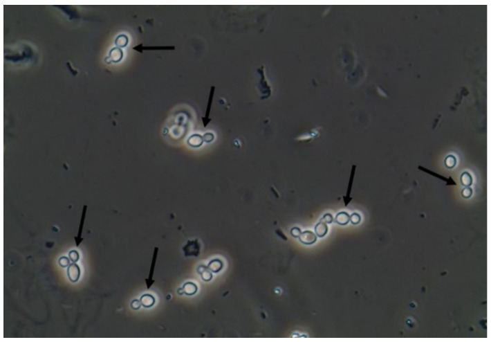
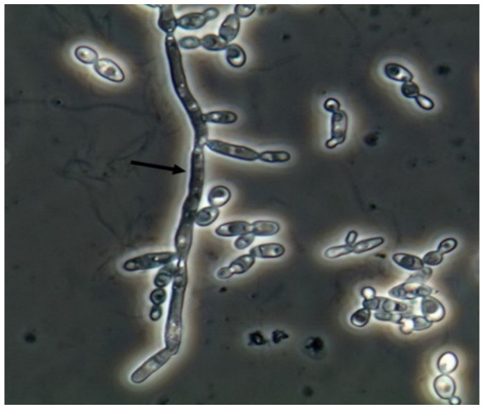
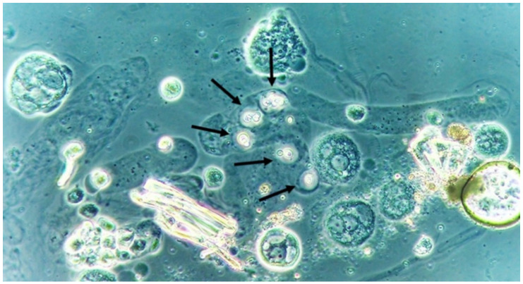
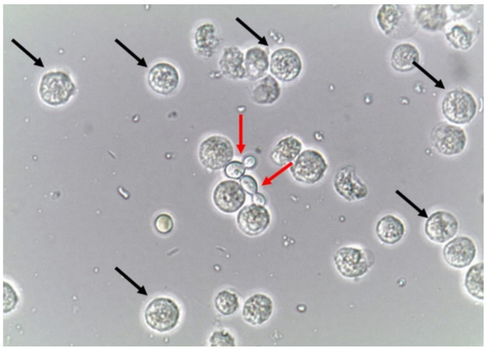
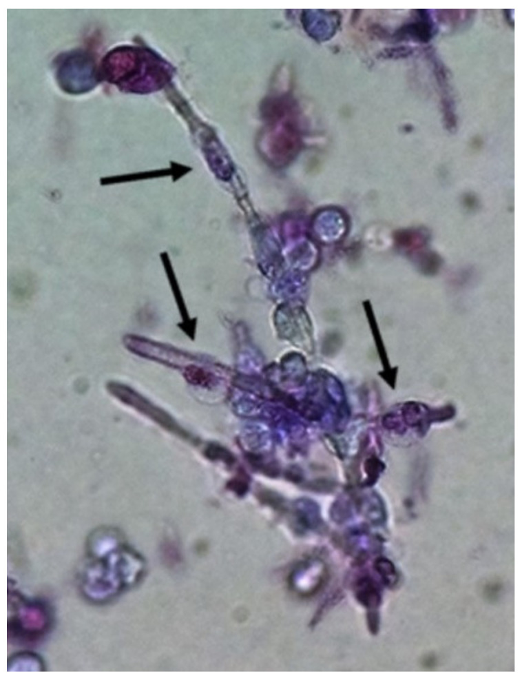
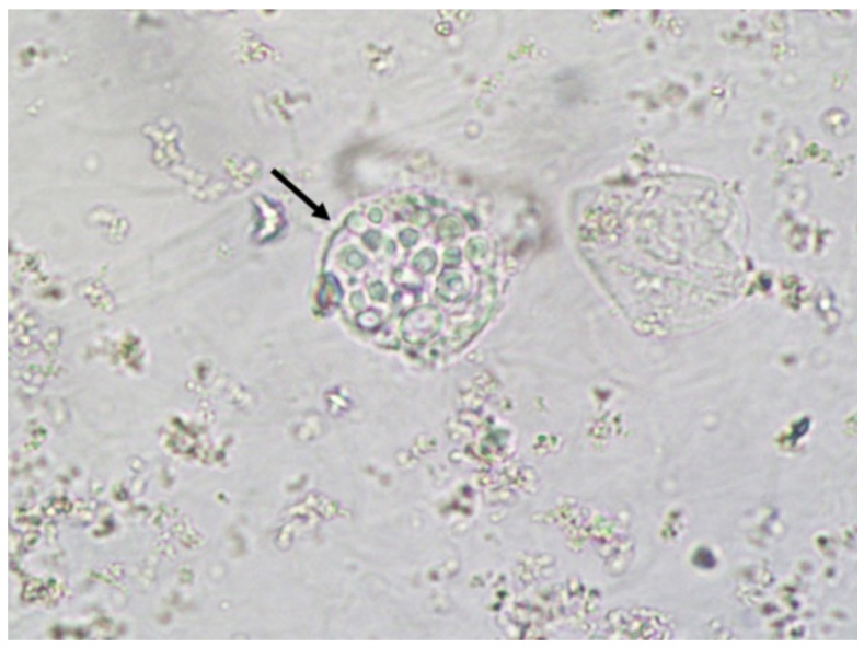
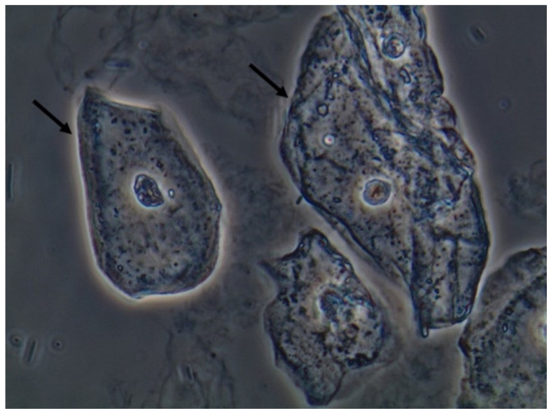
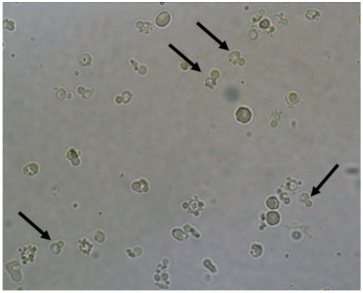
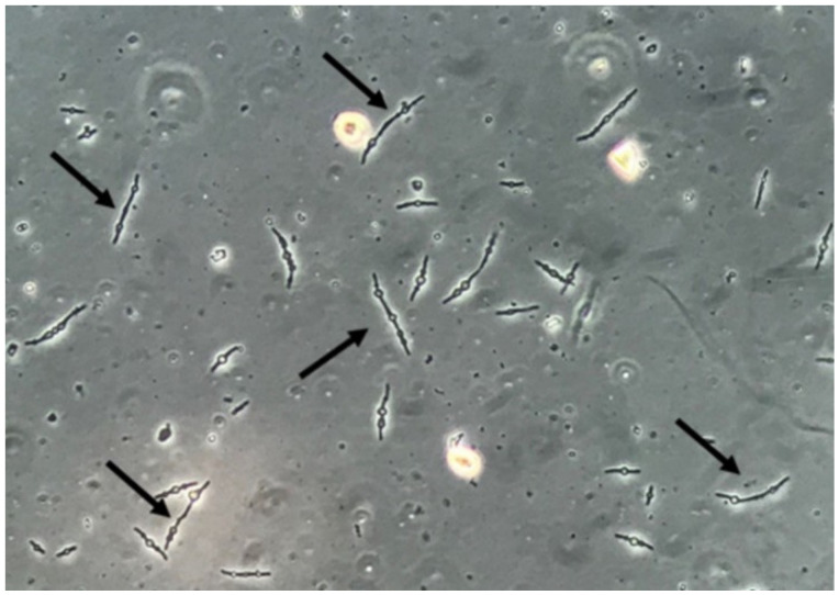
Fungi are pathogenic agents that can also cause disseminated infections involving the kidneys. Besides Candida, other agents like Cryptococcus spp. can cause urinary tract infection (UTI), as well as other non-yeast fungi, especially among immunocompromised patients. The detection and identification of fungi in urine samples (by microscopy and culture) plays an essential role in the diagnosis of fungal UTI. However, variable cutoff definitions and unreliable culture techniques may skew analysis of the incidence and outcome of candiduria. The sediment analysis plays a key role in the identification of fungal UTI because both yeasts and pseudohyphae are easily identified and can be used as a clinical sign of fungal UTI but should not be overinterpreted. Indeed, urine markers of the immune response (leukocytes), urine barriers of tissue protection (epithelial cells), and urine markers of kidney disease (urinary casts) can be found in urine samples. This work explores the manifestations associated with the fungal UTI from the urinalysis perspective, namely the urinary findings and clinical picture of patients with fungal UTI caused by Candida spp., aspects associated with the immune response, and the future perspectives of urinalysis in the diagnosis of this clinical condition.

Keywords: Candida spp.; fungal infection; immune response; urinary tract infection; urine sediment.


Conflict of interest statement

The authors declare no conflict of interest.

Figures

Figure 1
Figure 1
Candida albicans yeasts in the fresh and unstained urine sediment. Phase contrast microscopy. Original magnification: 400×. Courtesy: Controllab.
Figure 2
Figure 2
Candida albicans pseudohyphae in the fresh and unstained urine sediment. Phase contrast microscopy. Original magnification: 400×. Courtesy: Controllab.
Figure 3
Figure 3
Cryptococcus neoformans encapsuled yeasts within a urinary cast. Fresh and unstained urine sediment. Phase contrast microscopy. Original magnification: 400×.
Figure 4
Figure 4
Leukocytes (some of them pointed with black arrows) and yeast cells (red arrows). Fresh and unstained urine sediment. Bright field microscopy. Original magnification: 400×. Courtesy: Controllab.
Figure 5
Figure 5
Neutrophils (some of them pointed) firmly attached to Candida albicans pseudohyphae presumably trying to perform phagocytosis of the fungal structure. Urine sediment stained with Sternheimer-Malbin stain. Bright field microscopy. Original magnification: 400×.
Figure 6
Figure 6
Macrophage with Cryptococcus neoformans yeasts engulfed. Fresh and unstained urine sediment. Bright field microscopy. Original magnification: 400×.
Figure 7
Figure 7
Squamous epithelial cells in the fresh and unstained urine sediment. Phase contrast microscopy. Original magnification: 400×. Courtesy: Controllab.
Figure 8
Figure 8
Acanthocytes (some of them pointed). Fresh and unstained urine sediment. Bright field microscopy. Original magnification: 400×.
Figure 9
Figure 9
Multiresistant Escherichia coli bacteria forming filamentous forms and spheroplasts. Fresh and unstained urine sediment. Phase contrast microscopy. Original magnification: 400×.

References

    1. Gharanfoli A., Mahmoudi E., Torabizadeh R., Katiraee F., Faraji S. Isolation, characterization, and molecular identification of Candida species from urinary tract infections. Curr. Med. Mycol. 2019;5:33–36. doi: 10.18502/cmm.5.2.1159. - DOI - PMC - PubMed
    1. Kauffman C.A. Diagnosis and management of fungal urinary tract infection. Infect. Dis. Clin. N. Am. 2014;28:61–74. doi: 10.1016/j.idc.2013.09.004. - DOI - PubMed
    1. Reilly R., Perazella M.A. Nephrology in 30 Days. 2nd ed. Lange; New York, NY, USA: 2013.
    1. Fogazzi G.B. The Urinary Sediment—An Integrated View. 3rd ed. Elsevier; San Francisco, CA, USA: 2010.
    1. Fisher F.J., Kavanagh K., Sobel J.D., Kauffman C.A., Newman C.A. Candida Urinary Tract Infection: Pathogenesis. Clin. Infect. Dis. 2011;52(Suppl. S6):S437–S451. doi: 10.1093/cid/cir110. - DOI - PubMed

LinkOut - more resources