. 2020 Oct 24;10(11):1477.
doi: 10.3390/biom10111477.

Enhanced Enrichment of Medaka Ovarian Germline Stem Cells by a Combination of Density Gradient Centrifugation and Differential Plating

Affiliations

Enhanced Enrichment of Medaka Ovarian Germline Stem Cells by a Combination of Density Gradient Centrifugation and Differential Plating

Jun Hyung Ryu et al. Biomolecules. .

Abstract

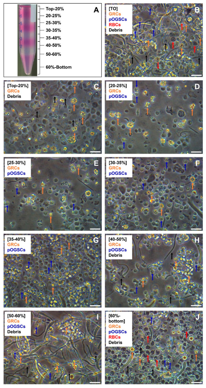
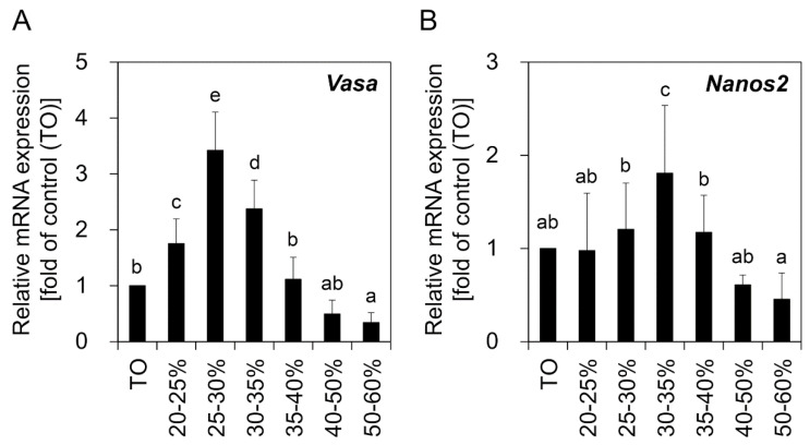
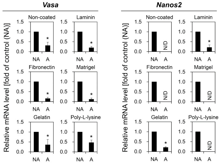
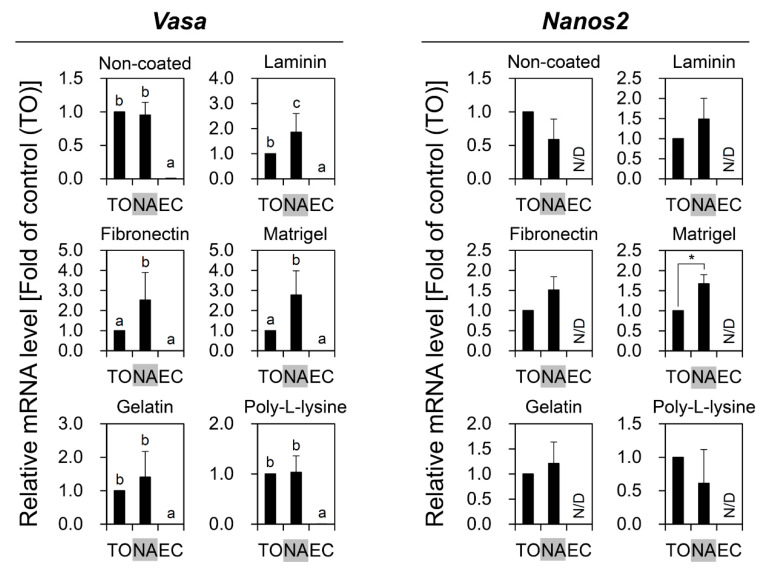
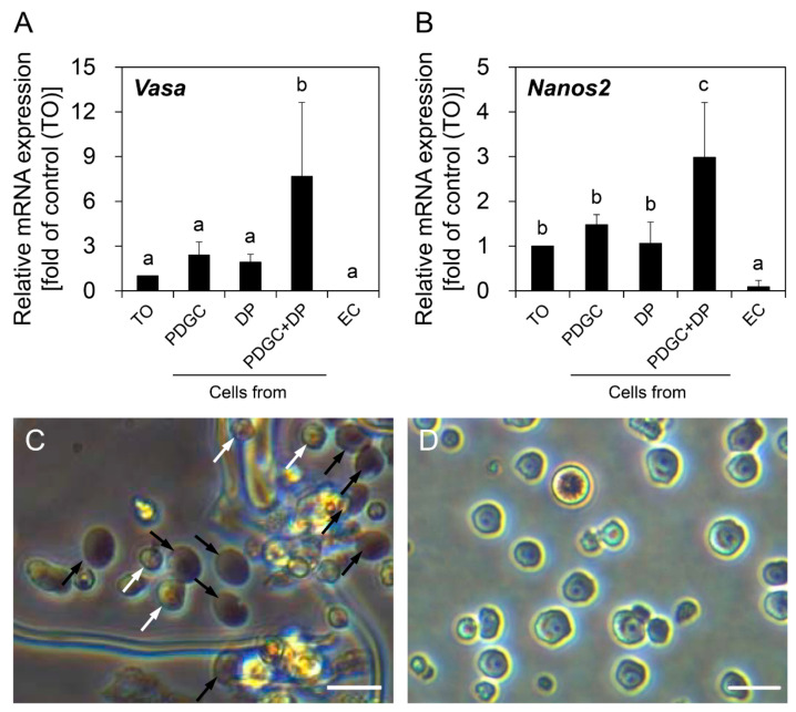
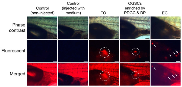
Fish ovarian germline stem cells (OGSCs) have great potential in various biological fields due to their ability to generate large numbers of mature eggs. Therefore, selective enrichment of OGSCs is a prerequisite for successful applications. To determine the optimal conditions for the enrichment of OGSCs from Japanese medaka (Oryzias latipes), we evaluated the effects of Percoll density gradient centrifugation (PDGC), differential plating (DP), and a combination of both methods. Based on cell morphology and gene expression of germ cell-specific Vasa and OGSC-specific Nanos2, we demonstrated that of seven density fractions obtained following PDGC, the 30-35% density fraction contained the highest proportion of OGSCs, and that Matrigel was the most effective biomolecule for the enrichment of Oryzias latipes OGSCs by DP in comparison to laminin, fibronectin, gelatin, and poly-l-lysine. Furthermore, we confirmed that PDGC and DP in combination significantly enhanced the efficiency of OGSC enrichment. The enriched cells were able to localize in the gonadal region at a higher efficiency compared to non-enriched ovarian cells when transplanted into the developing larvae. Our approach provides an efficient way to enrich OGSCs without using OGSC-specific surface markers or transgenic strains expressing OGSC-specific reporter proteins.

Keywords: Matrigel; density gradient centrifugation; differential plating; enrichment; fish; ovarian germline stem cells.


Conflict of interest statement

The authors declare no conflict of interest.

Figures

Figure 1
Figure 1
Separation of medaka (Oryzias latipes) crude ovarian cell populations by Percoll density gradient centrifugation (PDGC). Ovarian cells obtained from 10 adult females by mechanical and enzymatic dissociation were subjected to PDGC to separate the cells depending on their density. (A) Picture taken after PDGC. Density fractions from the top–20%, 20–25%, 25–30%, 30–35%, 35–40%, 40–50%, 50–60%, and 60%–bottom were collected separately, and the cells in each fraction were observed with an inverted microscope. (B) Image of the crude total ovarian cell population (TO) before cell separation. Granule-rich cells (GRCs), putative ovarian germline stem cells (pOGSCs), red blood cells (RBCs), and debris were observed. (C–J) Images of the cells from each density fraction after PDGC. pOGSCs were most abundant in 30-35% density fraction, and a lot of GRCs were identified in 35–60% density fractions. RBCs were concentrated in the 60%-bottom fraction, and tissue debris was abundant in the top–20% and 50–bottom fractions. Scale bar = 20 µm.
Figure 2
Figure 2
Relative expression of Vasa and Nanos2 genes in the ovary-derived cell populations separated by PDGC. Crude TO was harvested by enzymatic dissociation of the ovaries derived from 10 adult females. Following PDGC, cells from each density fraction were subjected to qRT-PCR analysis for comparison of Vasa (A) and Nanos2 (B) expression levels. TO was used as a control. Significant increases in Vasa expression were observed in the cells from the 20–35% density fractions. For Nanos2, the highest expression level was observed in cells from the 30–35% density fraction. The values are expressed as mean ±SD. a–e Different letters indicate significant differences; p < 0.05.
Figure 3
Figure 3
Comparison of Vasa and Nanos2 expression between non-adhesive (NA) and adhesive (A) cells after differential plating (DP). Crude TO from five adult females was subjected to DP on plates coated with different adhesion molecules including gelatin, fibronectin, laminin, Matrigel, and poly-l-lysine. NA and A cells were collected separately and analyzed by qRT-PCR to compare Vasa and Nanos2 expression levels. Regardless of the type of adhesion molecule used, the expression levels of both Vasa and Nanos2 were significantly higher in NA than A cells. The values are expressed as mean ± SD. Asterisks (*) indicate significant differences; p < 0.05. N/D, not detected.
Figure 4
Figure 4
Comparison of Vasa and Nanos2 expression between crude TO and NA cells derived from DP of TO. TO from five adult female fish was subjected to DP on plates coated with gelatin, fibronectin, laminin, Matrigel, or poly-l-lysine. NA cells were then collected from each plate. TO, NA, and Japanese medaka embryonic cells (EC), which were used as a negative control, were subjected to qRT-PCR for comparison of Vasa and Nanos2 expression levels. Significant increases in Vasa expression compared to TO were observed in NA cells from laminin, fibronectin, or Matrigel-coated plates. Only NA cells from Matrigel-coated plates showed significantly higher Nanos2 expression than TO. The values are expressed as mean ±SD. a-c Different letters and asterisk (*) indicate significant differences; p < 0.05. N/D, not detected.
Figure 5
Figure 5
Effects of combinatorial use of PDGC and DP on the enrichment of OGSCs. qRT-PCR analysis for Vasa (A) and Nanos2 (B) genes to evaluate the enrichment effect of the combinatorial use of PDGC and DP at the gene expression level. Crude TO was obtained from 10 adult females and separated by PDGC, Matrigel-based DP, or a combination of both. Cells from the 20–40% density fractions after PDGC and non-adherent cells after DP were used for analysis. In the combination of PDGC and DP, cells from the 20–40% density fractions after PDGC were subjected to Matrigel-based DP and the final non-adherent cells were used for analysis. Expression levels of Vasa and Nanos2 were significantly higher in the cells separated by PDGC and DP in combination compared to TO and the cells separated by PDGC or DP alone. Oryzias latipes EC were used as negative control. The values are expressed as mean ±SD. a–c Different letters indicate significant differences; p < 0.05. (C) Morphology of the cells separated by only DP. Putative OGSCs (white arrowheads) and red blood cells (black arrowheads) were observed with tissue debris. (D) Morphology of the cells separated by PDGC and DP in combination. Remarkable enrichment of putative OGSCs was observed. Scale bars = 20 µm.
Figure 6
Figure 6
Transplantation assay of OGSCs. Crude TO was obtained from 10 adult females and enriched by PDGC and DP in combination. TO, enriched OGSCs, or Oryzias latipes EC were transplanted into a peritoneal cavity of larvae at 11 dpf after labeling with PKH26. Localization of the donor cells in the gonadal region of the recipient larvae was examined at 20 dpf (9 days post-transplantation). The localization of the cells (dotted circles) was observed in the recipient larvae transplanted with TO and the cells enriched by PDGC and DP. The recipients transplanted with EC showed ectopic fluorescent signals (arrows) scattered in posterior abdominal regions. Scale bar = 100 µm.

Similar articles

Cited by

References

    1. Horan C.J., Williams S.A. Oocyte stem cells: Fact or fantasy? Reproduction. 2017;154:R23–R35. doi: 10.1530/REP-17-0008. - DOI - PubMed
    1. Nakamura S., Kobayashi K., Nishimura T., Higashijima S.-I., Tanaka M. Identification of Germline Stem Cells in the Ovary of the Teleost Medaka. Science. 2010;328:1561–1563. doi: 10.1126/science.1185473. - DOI - PubMed
    1. Yoshizaki G., Okutsu T., Morita T., Terasawa M., Yazawa R., Takeuchi Y. Biological Characteristics of Fish Germ Cells and their Application to Developmental Biotechnology. Reprod. Domest. Anim. 2012;47:187–192. doi: 10.1111/j.1439-0531.2012.02074.x. - DOI - PubMed
    1. Wong T.-T., Saito T., Crodian J., Collodi P. Zebrafish Germline Chimeras Produced by Transplantation of Ovarian Germ Cells into Sterile Host Larvae1. Biol. Reprod. 2011;84:1190–1197. doi: 10.1095/biolreprod.110.088427. - DOI - PMC - PubMed
    1. Yoshizaki G., Ichikawa M., Hayashi M., Iwasaki Y., Miwa M., Shikina S., Okutsu T. Sexual plasticity of ovarian germ cells in rainbow trout. Development. 2010;137:1227–1230. doi: 10.1242/dev.044982. - DOI - PubMed

Publication types

LinkOut - more resources