Review
. 2020 Oct 24;21(21):7901.
doi: 10.3390/ijms21217901.

Hijacking of Lipid Droplets by Hepatitis C, Dengue and Zika Viruses-From Viral Protein Moonlighting to Extracellular Release

Affiliations
Review

Hijacking of Lipid Droplets by Hepatitis C, Dengue and Zika Viruses-From Viral Protein Moonlighting to Extracellular Release

Alexandra P M Cloherty et al. Int J Mol Sci. .

Abstract

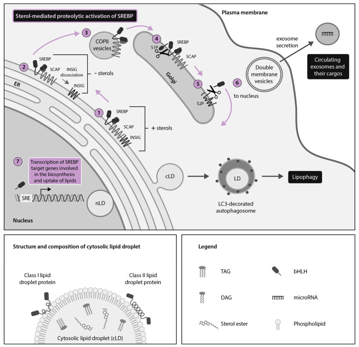
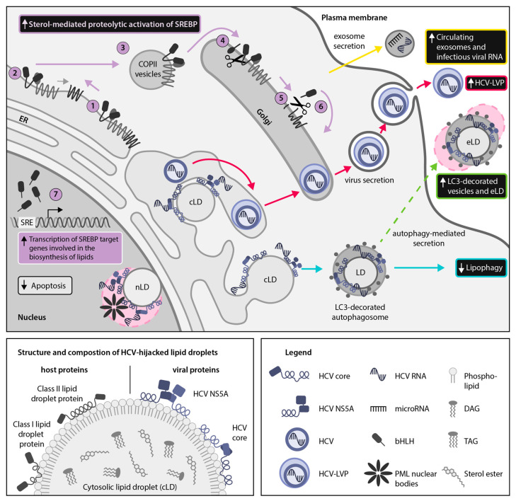
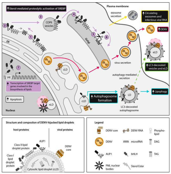
Hijacking and manipulation of host cell biosynthetic pathways by human enveloped viruses are essential for the viral lifecycle. Flaviviridae members, including hepatitis C, dengue and Zika viruses, extensively manipulate host lipid metabolism, underlining the importance of lipid droplets (LDs) in viral infection. LDs are dynamic cytoplasmic organelles that can act as sequestration platforms for a unique subset of host and viral proteins. Transient recruitment and mobilization of proteins to LDs during viral infection impacts host-cell biological properties, LD functionality and canonical protein functions. Notably, recent studies identified LDs in the nucleus and also identified that LDs are transported extracellularly via an autophagy-mediated mechanism, indicating a novel role for autophagy in Flaviviridae infections. These developments underline an unsuspected diversity and localization of LDs and potential moonlighting functions of LD-associated proteins during infection. This review summarizes recent breakthroughs concerning the LD hijacking activities of hepatitis C, dengue and Zika viruses and potential roles of cytoplasmic, nuclear and extracellular LD-associated viral proteins during infection.

Keywords: SREBP (sterol regulatory element-binding protein) pathway; Zika virus; autophagy; dengue virus; hepatitis C virus; lipid droplets.


Conflict of interest statement

The authors declare no conflict of interest.

Figures

Figure 1
Figure 1
The sterol regulatory element-binding protein (SREBP) pathway is a master regulator of cellular lipid droplet (LD) metabolism. LDs are spherical organelles composed of a phospholipid monolayer surrounding a core of neutral lipids. LDs have been described in both the cytoplasm (cLDs) and nucleus (nLDs) of homeostatic cells, with cLDs being better described. cLDs serve as docking platforms for Class I and Class II host LD proteins. LD metabolism is controlled by endoplasmic reticulum (ER)- and nuclear membrane-bound proteins called SREBPs, which control expression of key target genes involved in the biosynthesis and uptake of cholesterol and other lipids. When sterol levels are high, SREBPs are retained in an inactive state in the ER by SREBP-cleavage activating protein (SCAP) and insulin-induced gene (INSIG) (1). When sterol levels are low, INSIG dissociates from SCAP at the ER (2) and coat protein II (COPII)-coated vesicles transport SREBP-SCAP complexes to the Golgi apparatus (3), where the active N-terminal portion of SREBPs is cleaved by site-1 protease (S1P) (4) and site-2 protease (S2P) (5). The N-terminal portion of SREBPs then translocates to the nucleus (6) to act as a transcription factor coordinating expression of many lipid-metabolism related genes (7) that can subsequently impact LD biogenesis. SREBP regulation of LD metabolism can be further fine-tuned by exosome-packaged host microRNAs (miR) such as miR-29 (inhibits SCAP and SREBP-1 expression), miR-33 (inhibits SREBP-1 expression) and miR-24 (inhibits INSIG expression). SREBP-regulated LD homeostasis also intersects with other cellular pathways such as lipophagy, which catabolizes LDs in microtubule-associated protein light chain 3 (LC3)-decorated autophagosomes to liberate their component lipids.
Figure 2
Figure 2
Hepatitis C virus (HCV) hijacks sterol regulatory element-binding protein (SREBP)-regulated host lipid metabolism. HCV relies on manipulation of host lipid metabolism for virion packaging and release as lipid-coated lipoviroparticles (LVPs) (red arrows). Thus, HCV infection results in a multi-faceted disruption of lipid homeostasis, including remodeling of intracellular lipids into a membranous web, increased sterol-mediated SREBP activation and transcription of SREBP target genes (purple arrows: (17)), reduced autophagic catabolism of lipid droplets (LDs) (cyan arrows) and enhanced secretion of exosomes containing infectious viral RNA and host microRNAs that regulate lipid metabolism (yellow arrows). Here, we propose that the cytoplasmic LD (cLD)-associated HCV proteins core and non-structural protein 5A (NS5A) may also use nuclear LDs (nLDs) as a sequestration platform for interfering with host LD-associated proteins (pink circle). In the nucleus, LD-association of NS5A and core may facilitate their interaction with promyelocytic leukemia (PML) nuclear bodies, resulting in inhibition of host cell apoptosis. In addition, we hypothesize that hijacking of secreted microtubule-associated protein light chain 3 (LC3)-decorated vesicles containing LDs may be an additional mechanism for release of HCV virions, viral proteins or infectious viral RNA to escape the host cell (green arrow). Hypothetical pathways are highlighted with dotted lines.
Figure 3
Figure 3
Dengue virus (DENV) dysregulates host lipid metabolism. DENV perturbs host lipid homeostasis by enhancing sterol regulatory element-binding protein (SREBP) activation (purple arrows: (17)), leading to increased lipid droplet (LD) formation. DENV stimulates LD degradation via lipophagy by enhancing (ancient ubiquitous protein 1) AUP1 translocation from cytoplasmic LDs (cLDs) to microtubule-associated protein light chain 3 (LC3)-decorated autophagosomes (cyan arrows). Enhanced LD catabolism provides free fatty acids to serve as an energy source for production of infectious virions (red arrows). A recent report has shown that infectious viral RNA and extracellular LDs (eLDs) are contained within LC3-decorated extracellular vesicles in DENV-infected cells, suggesting that eLDs could be used as anchors for viral proteins. We hypothesize that the cLD-associated DENV core protein may also co-localize with eLDs in order to facilitate extracellular dissemination of viral RNA (green arrows). In addition, we propose that the DENV core protein may utilize nLDs as a scaffold for interacting with promyelocytic leukemia (PML) nuclear bodies (pink circle), thereby promoting host cell apoptosis and suppressing host interferon responses. Hypothetical pathways are highlighted with dotted lines.

References

    1. Tauchi-Sato K., Ozeki S., Houjou T., Taguchi R., Fujimoto T. The surface of lipid droplets is a phospholipid monolayer with a unique fatty acid composition. J. Biol. Chem. 2002;277:44507–44512. doi: 10.1074/jbc.M207712200. - DOI - PubMed
    1. Zweytick D., Athenstaedt K., Daum G. Intracellular lipid particles of eukaryotic cells. Biochim. Biophys. Acta-Rev. Biomembr. 2000;1469:101–120. doi: 10.1016/S0005-2736(00)00294-7. - DOI - PubMed
    1. Romanauska A., Köhler A. The inner nuclear membrane is a metabolically active territory that generates nuclear lipid droplets. Cell. 2018;174:700–715.e18. doi: 10.1016/j.cell.2018.05.047. - DOI - PMC - PubMed
    1. Hariri H., Rogers S., Ugrankar R., Liu Y.L., Feathers J.R., Henne W.M. Lipid droplet biogenesis is spatially coordinated at ER–vacuole contacts under nutritional stress. EMBO Rep. 2018;19:57–72. doi: 10.15252/embr.201744815. - DOI - PMC - PubMed
    1. Wu Y.W., Mettling C., Wu S.R., Yu C.Y., Perng G.C., Lin Y.S., Lin Y.L. Autophagy-associated dengue vesicles promote viral transmission avoiding antibody neutralization. Sci. Rep. 2016;6:1–10. doi: 10.1038/srep32243. - DOI - PMC - PubMed