

Influence of Polyethylene Glycol on Leaf Anatomy, Stomatal Behavior, Water Loss, and Some Physiological Traits of Date Palm Plantlets Grown In Vitro and Ex Vitro
- PMID: 33114523
- PMCID: PMC7693573
- DOI: 10.3390/plants9111440
Influence of Polyethylene Glycol on Leaf Anatomy, Stomatal Behavior, Water Loss, and Some Physiological Traits of Date Palm Plantlets Grown In Vitro and Ex Vitro
Abstract
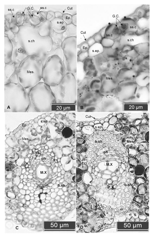
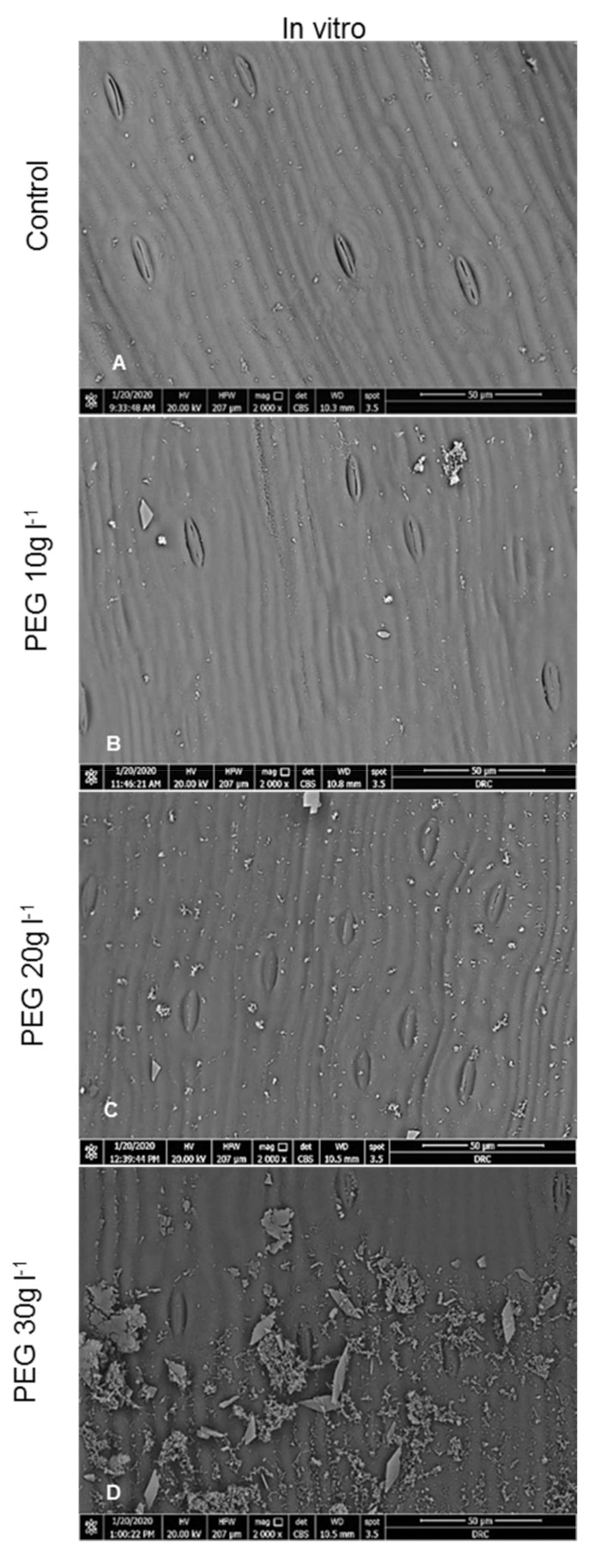
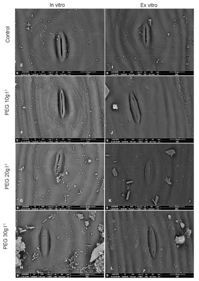
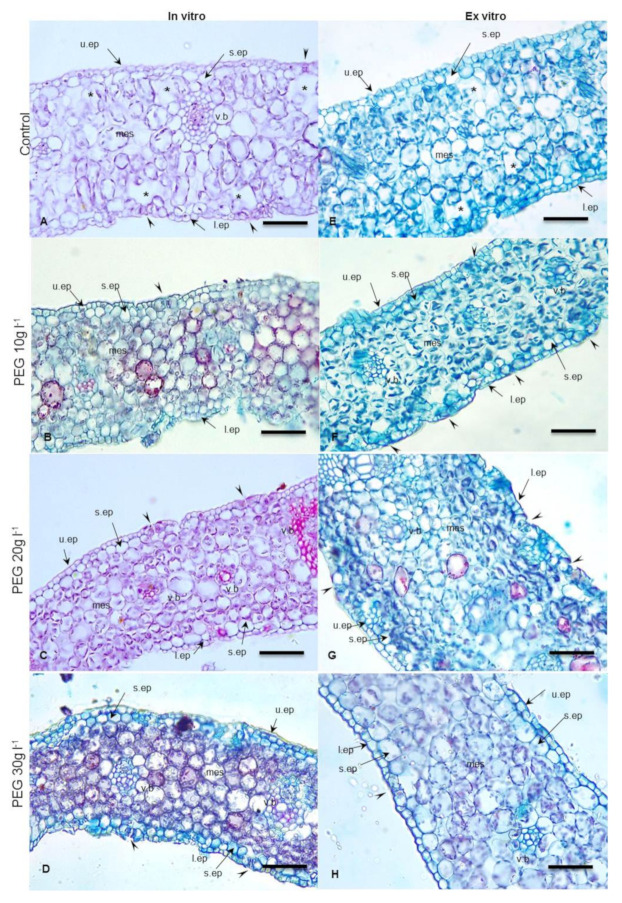
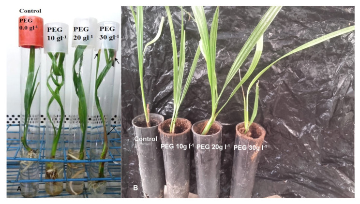
Few reports explain the mechanism of PEG action on stomatal behavior and anatomical structure and analyze the photosynthetic pigments of in vitro date palm plantlets for better tolerance to ex vitro exposure. The main challenge for in vitro micropropagation of date palm techniques remains restricted to high survival rates and vigorous growth after ex vitro transplantation. In vitro hardening is induced by Polyethylene glycol PEG (0.0, 10, 20, 30 g L-1) for 4 weeks. Leaf anatomy, stomatal behavior, water loss %, photosynthetic pigments, and reducing sugars were examined in date palm plantlets (Phoenix dactylifera L.) cv. (Sewi) after 4 weeks from in vitro PEG treatment and after 4 weeks from ex vitro transplanting to the greenhouse. Leaf anatomy and the surface ultrastructure of in vitro untreated leaves showed a thin cuticle layer, wide opened malfunctioning stomata, and abnormal leaf anatomy. Furthermore, addition of PEG resulted in increasing cuticle thickness, epicuticular wax depositions, and plastids density, improving the stomatal ability to close and decreasing the stomatal aperture length while reducing the substomatal chambers and intercellular spaces in the mesophyll. As a result, a significant reduction in water loss % was observed in both in vitro and ex vitro PEG treated leaves as compared to untreated ones, which exhibited rapid wilting when exposed to low humidity for 4 h. PEG application significantly increased Chlorophylls a, b and carotenoids concentrations, especially 10, 20 g L-1 treatments, which were sequentially reflected in increasing the reducing sugar concentration. However, leaves of plantlets treated with PEG at 30 g L-1 became yellow and had necrosis ends with death. In vitro hardening by 20 g L-1 PEG increased the survival rate of plantlets to 90% after ex vitro transfer compared to 63% recorded for the untreated plantlets. Therefore, this application provides normal date palm plantlets developed faster and enhances survival after ex vitro transfer.
Keywords: Phoenix dactylifera; ex vitro transfer; in vitro hardening; leaf anatomy; polyethylene glycol (PEG); stomata; survival; water loss %.
Conflict of interest statement
The authors declare no conflict of interest.
Figures
References
-
- Al-Khayri J. Date palm Phoenix dactylifera L. micropropagation. In: Jain S., Haggman H., editors. Protocols for Micropropagation of Woody Trees and Fruits. Springer; Berlin/Heidelberg, Germany: 2007.
-
- Abd El Bar O.H.A., El Dawayati M.M. Histological changes on regeneration in vitro culture of date palm (Phoenix dactylifera) leaf explants. Aust. J. Crop Sci. 2014;8:848–855.
-
- Al-Khayri J.M. In vitro germination of somatic embryos in date palm: Effect of auxin concentration and strength of MS salts. Curr. Sci. 2003;84:680–683.
-
- Quiroz-Figueroa F.R., Rojas-Herrera R., Galaz-Avalos R.M., Loyola-Vargas V.M. Embryo production through somatic embryogenesis can be used to study cell differentiation in plants. Plant Cell Tissue Organ Cult. 2006;86:285. doi: 10.1007/s11240-006-9139-6. - DOI
-
- Al Kaabi H., Rhiss A., Hassan M. Effect of auxins and cytokinins on the in vitro production of date palm bud generative tissues and on the number of differentiated buds; Proceedings of the Second International Conference on Date Palms; Al Ain, UAE. 25–27 March 2001; pp. 47–86.
Grants and funding
LinkOut - more resources
Full Text Sources
Research Materials
