. 2020 Oct 26;25(21):4940.
doi: 10.3390/molecules25214940.

Synthetic Makaluvamine Analogs Decrease c-Kit Expression and Are Cytotoxic to Neuroendocrine Tumor Cells

Affiliations

Synthetic Makaluvamine Analogs Decrease c-Kit Expression and Are Cytotoxic to Neuroendocrine Tumor Cells

Zviadi Aburjania et al. Molecules. .

Abstract

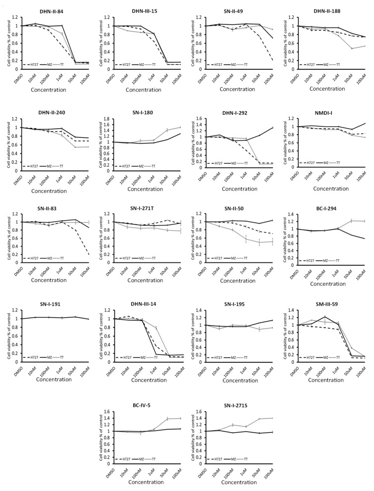
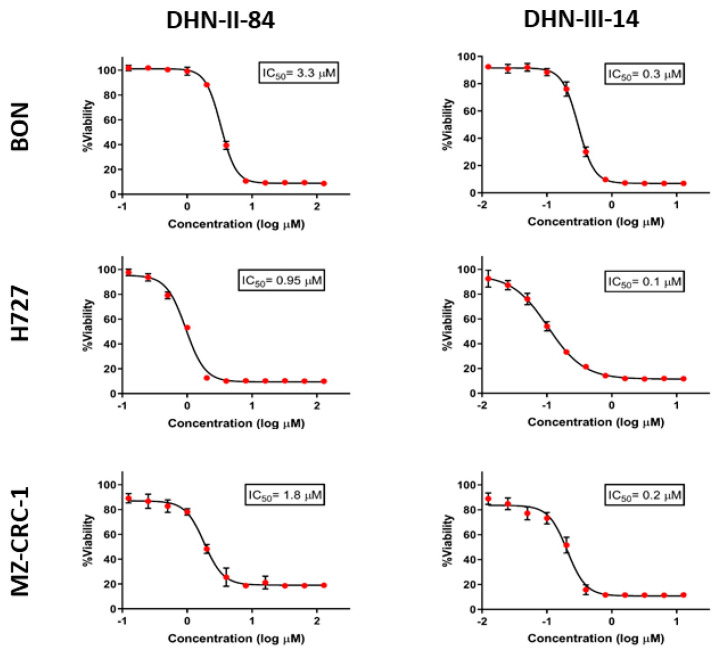
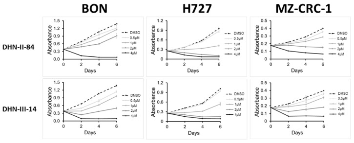
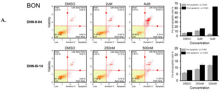
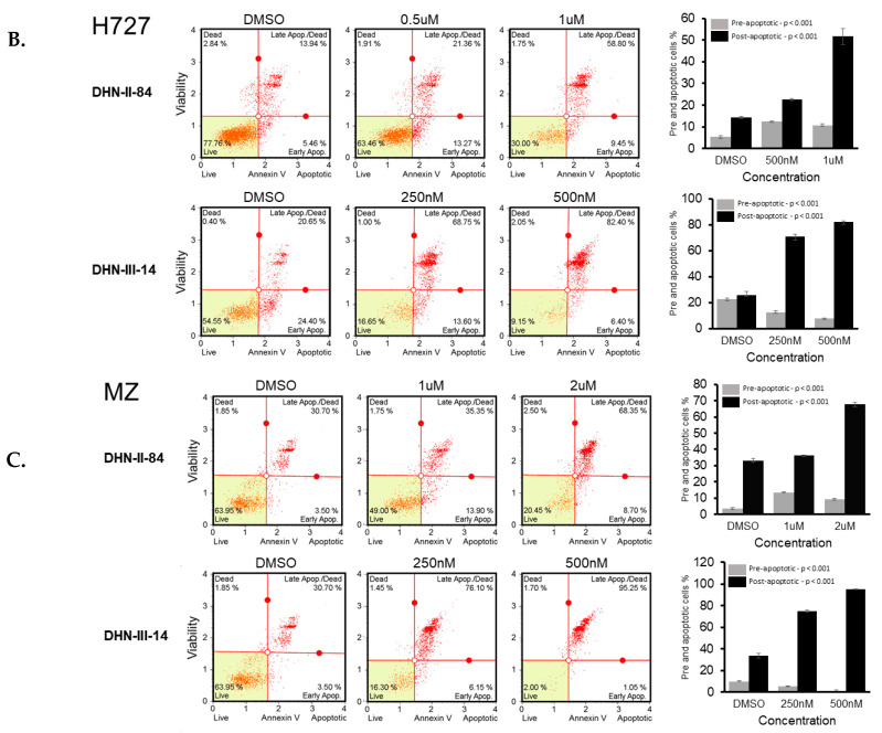
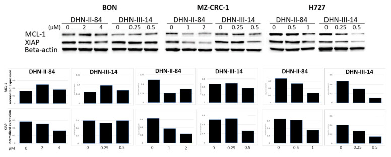
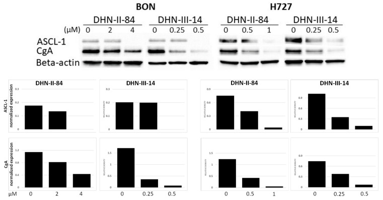
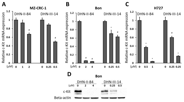
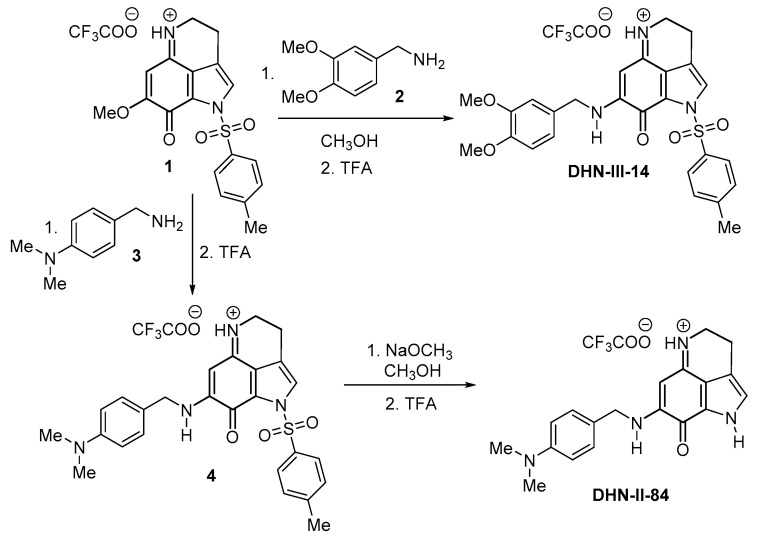
In an effort to discover viable systemic chemotherapeutic agents for neuroendocrine tumors (NETs), we screened a small library of 18 drug-like compounds obtained from the Velu lab against pulmonary (H727) and thyroid (MZ-CRC-1 and TT) neuroendocrine tumor-derived cell lines. Two potent lead compounds (DHN-II-84 and DHN-III-14) identified from this screening were found to be analogs of the natural product makaluvamine. We further characterized the antitumor activities of these two compounds using pulmonary (H727), thyroid (MZ-CRC-1) and pancreatic (BON) neuroendocrine tumor cell lines. Flow cytometry showed a dose-dependent increase in apoptosis in all cell lines. Induction of apoptosis with these compounds was also supported by the decrease in myeloid cell leukemia-1 (MCL-1) and X-chromosome linked inhibitor of apoptosis (XIAP) detected by Western blot. Compound treatment decreased NET markers chromogranin A (CgA) and achaete-scute homolog 1 (ASCL1) in a dose-dependent manner. Moreover, the gene expression analysis showed that the compound treatment reduced c-Kit proto-oncogene expression in the NET cell lines. Induction of apoptosis could also have been caused by the inhibition of c-Kit expression, in addition to the known mechanisms such as damage of DNA by topoisomerase II inhibition for this class of compounds. In summary, makaluvamine analogs DHN-II-84 and DHN-III-14 induced apoptosis, decreased neuroendocrine tumor markers, and showed promising antitumor activity in pulmonary, thyroid, and pancreatic NET cell lines, and hold potential to be developed as an effective treatment to combat neuroendocrine tumors.

Keywords: NET; alkaloid; cancer; makaluvamine; neuroendocrine; therapeutic; tumor.


Conflict of interest statement

The authors declare no conflict of interest. The funding agencies had no role in the design, execution, interpretation, or on the preparation of this manuscript.

Figures

Figure 1
Figure 1
Chemical structures of makaluvamine A, makaluvamine D and the test compounds DHN-II-84 and DHN-III-14.
Figure 2
Figure 2
Chemical structures of 18 compounds used in the preliminary screening against pulmonary (H727) and thyroid (MZ-CRC-1 and TT) neuroendocrine tumor cell lines.
Figure 3
Figure 3
Test compounds showed varying degrees of cytotoxic effects on neuroendocrine cell lines: H727, MZ-CRC-1 and TT in MTT assay. Experiments were performed in quadruplicates and data are plotted as mean ± SEM. Prominent effects were displayed by DHN-II-84 and DHN-III-14.
Figure 4
Figure 4
The IC50 values for DHN-II-84 and DHN-III-14 determined by CellTiter-Glo cell cytotoxicity assay against neuroendocrine cell lines H727, MZ-CRC-1 and BON. Experiments were performed in quadruplicates and data are plotted as mean ± SEM.
Figure 5
Figure 5
DNH-II-84 and DNH-III-14 inhibit neuroendocrine cell proliferation. BON, H727 and MZ-CRC-1 cell lines were treated with increasing concentrations of DNH-II-84 and DNH-III-14 (0–4 µM). Absorbance was measured on day 0, 2, 4 and 6 using MTT assay. Experiments were performed in quadruplicates and data are plotted as mean ± SEM.
Figure 6
Figure 6
DHN-II-84 and DHN-III-14 increase apoptosis in neuroendocrine cancer cells. BON (A), H727 (B) and MZ-CRC-1 (C) cell lines were treated with increasing doses of DHN-II-84 and DNH-III-14, with half-maximum inhibitory concentration being the highest dose. A dose-dependent increase in apoptosis was observed in all cell lines. The highest response to the drug treatment was seen in MZ-CRC-1 when treated with DHN-III-14 (C), and the lowest response was seen with BON treated with DHN-II-14 (A). Bar graphs represent the mean of pre- and post-apoptotic cells ± SEM. Measurements were performed in triplicates. Statistical differences of pre- and post-apoptotic cells after treatment were determined by one-way ANOVA.
Figure 6
Figure 6
DHN-II-84 and DHN-III-14 increase apoptosis in neuroendocrine cancer cells. BON (A), H727 (B) and MZ-CRC-1 (C) cell lines were treated with increasing doses of DHN-II-84 and DNH-III-14, with half-maximum inhibitory concentration being the highest dose. A dose-dependent increase in apoptosis was observed in all cell lines. The highest response to the drug treatment was seen in MZ-CRC-1 when treated with DHN-III-14 (C), and the lowest response was seen with BON treated with DHN-II-14 (A). Bar graphs represent the mean of pre- and post-apoptotic cells ± SEM. Measurements were performed in triplicates. Statistical differences of pre- and post-apoptotic cells after treatment were determined by one-way ANOVA.
Figure 7
Figure 7
DHN-II-84 and DHN-III-14 decrease antiapoptotic markers in neuroendocrine cancer cells. Protein levels of MCL-1 and XIAP were evaluated by Western blotting after 48 h of treatment. MCL-1, myeloid cell leukemia-1; XIAP, X-linked inhibitor of apoptosis protein. Both antiapoptotic markers decreased in MZ-CRC-1 and H727 cell lines. Only DHN-II-84 treatment in BON cell line decreased XIAP. Densitometry graphs of MCL-1 and XIAP protein levels normalized to beta-actin band intensity levels. Relative density levels shown after treatment.
Figure 8
Figure 8
DHN-II-84 and DHN-III-14 decrease neuroendocrine tumor markers in cancer cell lines. A dose-dependent decrease in neuroendocrine tumor markers was observed in BON and H727 cell lines. ASCL-1, achaete-scute homolog 1; CgA, chromogranin A. Protein bands normalized to beta-actin. Relative protein band intensities shown after treatment.
Figure 9
Figure 9
DHN-II-84 and DHN-III-14 decrease c-Kit expression in MZ-CRC-1 (A), BON (B) and H727 (C) cells. The mRNA level of c-Kit was quantified by RT-qPCR in the cell lines following the treatment with DNH-II-84 and DNH-III-14 with half-IC50 and IC50 concentrations. The expression levels of mRNA are plotted relative to no treatment. All values are presented as mean relative fold ± SEM (* p < 0.05 and # p < 0.01). (D) Western blot demonstrating a basal expression level of c-Kit protein in BON cells and its reduction with DHN-II-84 and DHN-III-14 treatment.
Figure 10
Figure 10
Synthesis of test compounds DHN-II-84 and DHN-III-14.

References

    1. Siegel R., Ma J., Zou Z., Jemal A. Cancer statistics, 2014. CA Cancer J. Clin. 2014;64:9–29. doi: 10.3322/caac.21208. - DOI - PubMed
    1. Yao J.C., Hassan M., Phan A., Dagohoy C., Leary C., Mares J.E., Abdalla E.K., Fleming J.B., Vauthey J.-N., Rashid A., et al. One Hundred Years after “Carcinoid”: Epidemiology of and Prognostic Factors for Neuroendocrine Tumors in 35,825 Cases in the United States. J. Clin. Oncol. 2008;26:3063–3072. doi: 10.1200/JCO.2007.15.4377. - DOI - PubMed
    1. Hauso O., Gustafsson B.I., Kidd M., Waldum H.L., Drozdov I., Chan A.K.C., Modlin I.M. Neuroendocrine tumor epidemiology. Cancer. 2008;113:2655–2664. doi: 10.1002/cncr.23883. - DOI - PubMed
    1. Cayo M., Greenblatt D., Kunnimalaiyaan M., Chen H. Carcinoid Tumors. Atlas Genet. Cytogenet. Oncol. Haematol. 2011;13:1255–1269. doi: 10.4267/2042/44440. - DOI
    1. Zandee W.T., Kamp K., Van Adrichem R.C., Feelders R.A., De Herder W.W. Effect of hormone secretory syndromes on neuroendocrine tumor prognosis. Endocr. Relat. Cancer. 2017;24:R261–R274. doi: 10.1530/ERC-16-0538. - DOI - PubMed

MeSH terms