Review
. 2020 Oct 26;11(11):1262.
doi: 10.3390/genes11111262.

Transcription Factors of the bHLH Family Delineate Vertebrate Landmarks in the Nervous System of a Simple Chordate

Affiliations
Review

Transcription Factors of the bHLH Family Delineate Vertebrate Landmarks in the Nervous System of a Simple Chordate

Lenny J Negrón-Piñeiro et al. Genes (Basel). .

Abstract

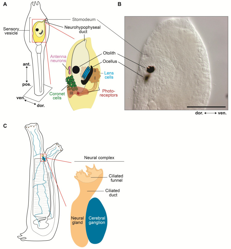
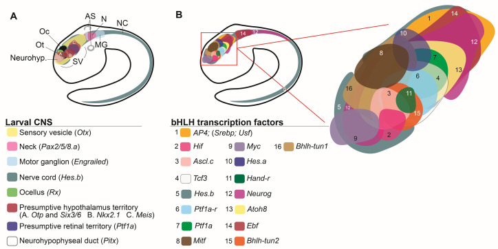
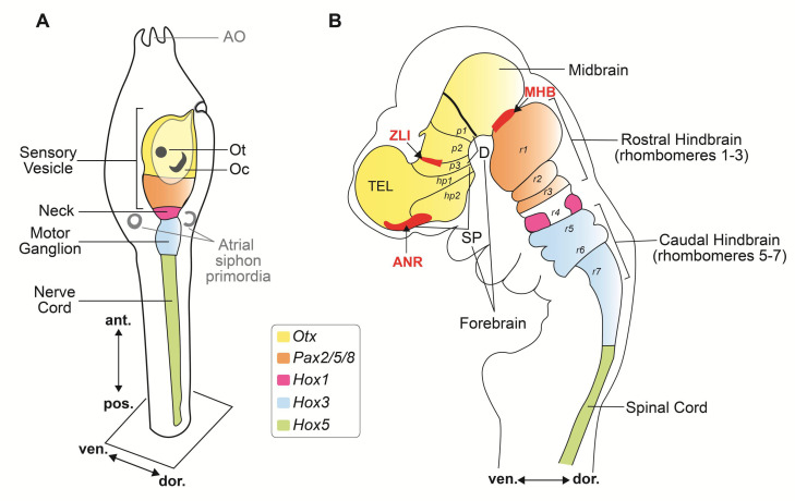
Tunicates are marine invertebrates whose tadpole-like larvae feature a highly simplified version of the chordate body plan. Similar to their distant vertebrate relatives, tunicate larvae develop a regionalized central nervous system and form distinct neural structures, which include a rostral sensory vesicle, a motor ganglion, and a caudal nerve cord. The sensory vesicle contains a photoreceptive complex and a statocyst, and based on the comparable expression patterns of evolutionarily conserved marker genes, it is believed to include proto-hypothalamic and proto-retinal territories. The evolutionarily conserved molecular fingerprints of these landmarks of the vertebrate brain consist of genes encoding for different transcription factors, and of the gene batteries that they control, and include several members of the bHLH family. Here we review the complement of bHLH genes present in the streamlined genome of the tunicate Ciona robusta and their current classification, and summarize recent studies on proneural bHLH transcription factors and their expression territories. We discuss the possible roles of bHLH genes in establishing the molecular compartmentalization of the enticing nervous system of this unassuming chordate.

Keywords: CNS; Ciona; ascidian; bHLH; epiphysis; hypophysis; hypothalamus; nervous system; notochord; sensory vesicle.


Conflict of interest statement

The authors declare no conflict of interest.

Figures

Figure 2
Figure 2
Larval sensory vesicle and adult neural complex of Ciona. (A) Schematic view of the sensory vesicle, the ‘brain’ of the ascidian larva, its sensory organs, and the primordia of the hypophysis and stomodeum. On the right side of the sensory vesicle reside the pigmented ocellus and the associated lens cells and photoreceptors (group I and II), whereas the left side contains the otolith, antenna neurons, coronet cells, and photoreceptor cells (group III). (B) Microphotograph of the trunk of a Ciona larva, showing the developing stomodeum, the otolith, and the ocellus. Scale bar: 25 µm. (C) Schematic view of the sessile filter-feeder adult, highlighting the neural complex, located between the two siphons, and its components, the cerebral ganglion and the neural gland. Nerve fibers from the neural complex (blue) innervate multiple organs and tissues. Adapted from [48,85,86].
Figure 3
Figure 3
The panoply of bHLH genes expressed in the modest CNS of the Ciona larva. (A) The four main subdivisions of the larval CNS, as delineated by the expression patterns of Otx (sensory vesicle) Pax2/5/8.a (neck), Engrailed (motor ganglion), and Hes.b (expressed in all previous subdivisions, and in the nerve cord). The region of the sensory vesicle posited to represent a proto-hypothalamus-retinal territory is delineated by the expression of Otp, Six3/6, Nkx2.1, Meis, and Ptf1a. The territory that gives rise to the photoreceptors associated to the ocellus is marked by the expression of Rx, and the neurohypophyseal primordium is labeled by the expression of Pitx. Adapted from [62]. (B) The palette of bHLH genes expressed in the Ciona SV and the complex tapestry that they delineate within this simple chordate brain. Each electronic brushstroke symbolizes the approximate expression pattern of a different bHLH gene, reconstructed from published WMISH expression analysis at mid/late-tailbud stage, according to the color/number code below this panel. The expression territories of Tcf3, Id.b and Mnt-r have not been included because the results of their respective WMISH were unclear. Adapted from: [29,50,84,108,120,145,163,164,165,166]. Abbreviations: AS, atrial siphon primordium; MG, motor ganglion; N, neck; NC, nerve cord; Neurohyp., neurohypophyseal primordium; Oc, ocellus; Ot, otolith; SV, sensory vesicle.
Figure 1
Figure 1
General organization and presumed homologous regions of the central nervous system (CNS) in ascidian larvae and vertebrate embryos. Drawings of a Ciona larva (A), ~18 h after fertilization and a mouse embryo (B), ~9 days old (stage E9.5). The developing nervous systems of these divergent chordates exhibit a comparable anterior-posterior sequential expression of the genes Otx (yellow), Pax2/5/8 (orange), Hox1 (magenta), Hox3 (blue), and Hox5 (green). Abbreviations: ANR, anterior neural ridge; ant., anterior; AO, adhesive organ; D, diencephalon; dor., dorsal; hp, hypothalamic prosomere; MHB, mid-hindbrain boundary; Oc, ocellus; Ot, otolith; p, prosomere; pos., posterior; r, rhombomere; SP, secondary prosencephalon; TEL, telencephalon; ven., ventral; ZLI, zona limitans intrathalamica. Adapted from [10,17,55].

References

    1. Delsuc F., Brinkmann H., Chourrout D., Philippe H. Tunicates and not cephalochordates are the closest living relatives of vertebrates. Nature. 2006;439:965–968. doi: 10.1038/nature04336. - DOI - PubMed
    1. Chabry L. Contribution a l’embryologie normale et teratologique des Ascidies simples. J. Anat. Physiol. (Paris) 1887;23:167–319.
    1. Conklin E.G. The organization and cell-lineage of the ascidian egg. Acad. Nat. Sci. :1905. doi: 10.5962/bhl.title.4801. - DOI
    1. Lawrence P.A., Levine M. Mosaic and regulative development: Two faces of one coin. Curr. Biol. 2006;16:R236–R239. doi: 10.1016/j.cub.2006.03.016. - DOI - PubMed
    1. Ortolani G. Risultati definitivi sulla distribuzione dei territory presuntivi degli organi nel germe di Ascidie allo stadio VIII, determinati con le marche al carbone. Section II: History and Philosophy of the Life Sciences. Pubbl. Stn. Zool. Napoli. 1954;25:161–187.

Publication types

MeSH terms

Substances

LinkOut - more resources