Review
. 2020 Oct 26;10(11):1485.
doi: 10.3390/biom10111485.

VDAC1 at the Intersection of Cell Metabolism, Apoptosis, and Diseases

Affiliations
Review

VDAC1 at the Intersection of Cell Metabolism, Apoptosis, and Diseases

Varda Shoshan-Barmatz et al. Biomolecules. .

Abstract

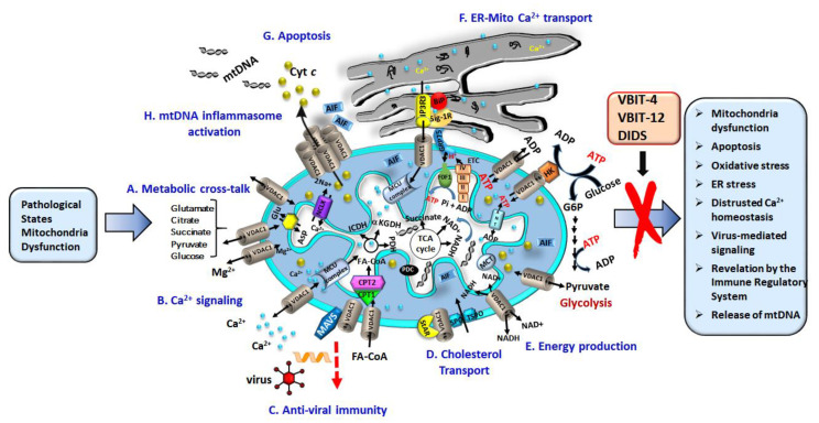
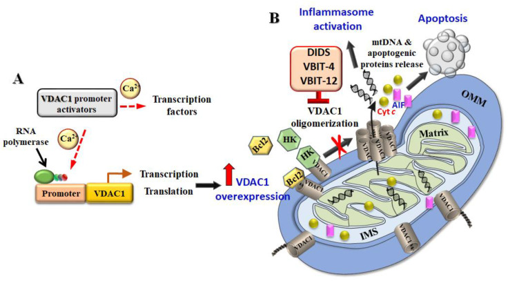
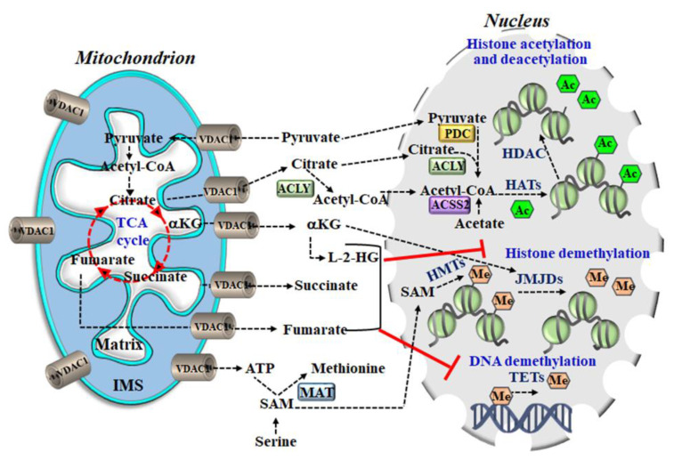
The voltage-dependent anion channel 1 (VDAC1) protein, is an important regulator of mitochondrial function, and serves as a mitochondrial gatekeeper, with responsibility for cellular fate. In addition to control over energy sources and metabolism, the protein also regulates epigenomic elements and apoptosis via mediating the release of apoptotic proteins from the mitochondria. Apoptotic and pathological conditions, as well as certain viruses, induce cell death by inducing VDAC1 overexpression leading to oligomerization, and the formation of a large channel within the VDAC1 homo-oligomer. This then permits the release of pro-apoptotic proteins from the mitochondria and subsequent apoptosis. Mitochondrial DNA can also be released through this channel, which triggers type-Ι interferon responses. VDAC1 also participates in endoplasmic reticulum (ER)-mitochondria cross-talk, and in the regulation of autophagy, and inflammation. Its location in the outer mitochondrial membrane, makes VDAC1 ideally placed to interact with over 100 proteins, and to orchestrate the interaction of mitochondrial and cellular activities through a number of signaling pathways. Here, we provide insights into the multiple functions of VDAC1 and describe its involvement in several diseases, which demonstrate the potential of this protein as a druggable target in a wide variety of pathologies, including cancer.

Keywords: VDAC1; apoptosis; cancer; diseases; metabolism; mitochondria; virus.


Conflict of interest statement

The authors declare no conflict of interest

Figures

Figure 1
Figure 1
Voltage-dependent anion channel 1 (VDAC1) as a multi-functional channel mediates metabolites, nucleotides, and Ca2+ transport, controlling energy production, endoplasmic reticulum (ER)-mitochondria cross-talk, and apoptosis. VDAC1 is responsible for a number of functions in the cell and mitochondria including: (A) transfer of metabolites between the mitochondria and cytosol; (B) passage of Ca2+ to and from the intermembrane space (IMS) to facilitate Ca2+ signaling; (C) Mitochondrial antiviral-signaling protein (MAVS) associated with VDAC1 enable anti-viral signaling. (D) Transfer of acetyl coenzyme-A (acyl-CoAs) across the outer mitochondrial membrane (OMM) to the IMS, for conversion into acylcarnitine by CPT1a for further processing by β-oxidation. Together with Star and translocator protein (TSPO), VDAC1 forms the multi-protein transduceosome, which transports cholesterol. (E) Recycling ATP/ADP, NAD+/NADH, and acyl-CoA between the cytosol and the IMS, and regulating glycolysis via association with HK; (F) contributing to ER-mitochondria contacts, where Ca2+ released by IP3 activation of inositol 1,4,5-trisphosphate receptors (IP3R) in the ER is directly transferred to IMS via VDAC1, and then is transported to the matrix by the Ca2+ uniporter (MCU complex). In the matrix Ca2+ regulates energy production via activation of the tricarboxylic acid cycle (TCA) cycle enzymes: pyruvate dehydrogenase (PDH), isocitrate dehydrogenase (ICDH), and α-ketoglutarate dehydrogenase (α-KGDH). The electron transport chain (ETC) and the ATP synthase (FoF1) are also presented. (G) VDAC1 oligomers forming a hydrophilic protein-conducting channel capable of mediating the release of apoptogenic proteins (e.g., Cyto c and apoptosis-inducing factor (AIF)) from the mitochondrial IMS to the cytosol, leading to apoptosis. (H). VDAC1 oligomers allow mtDNA release triggering inflammasome activation. Pathological conations lead to dysfunction of the mitochondria as reflected in the activities presented in the box on the right. These altered activities can be prevented by VDAC1-interacting molecules, such as DIDS, VBIT-4 and VBIT-12.
Figure 2
Figure 2
Sequence of events leading to VDAC1 overexpression, oligomerization, and apoptosis. Apoptosis stimuli or pathological conditions enhance VDAC1 expression via Ca2+ or transcription factors (TFs) activating promoter, leading to VDAC1 transcription.
Figure 3
Figure 3
Proposed model for the mechanism of action for activators of the VDAC1 promoter leading oligomerization, and apoptosis. (A) Apoptotic stimuli act either by directly activating the VDAC1 promoter or by stimulating the activity of TFs, thereby inducing VDAC1. Ca2+ directly or via a TF leads to promoter activation. (B) Overexpressed VDAC1 shifts the equilibrium to the VDAC1 oligomeric state, mediating the release of apoptogenic proteins, and leading to apoptosis. VDAC1-interacting molecules such as VBIT-4, inhibit VDAC1 oligomerization and apoptosis.
Figure 4
Figure 4
Mitochondrial metabolites are transported by VDAC1 to the cytosol and are then transported to the nucleus where they serve as substrates for enzymes that modify chromatin. Mitochondrial metabolic pathways including the TCA cycle generate substrates required for the methylation, acetylation, or demethylation reactions that modify chromatin. Specifically, histone acetylation by histone acetyltransferases (HATs) is dependent on the availability of acetyl groups provided by acetyl-CoA, which is produced in the cytosol by ACLY using citrate exported from the TCA cycle in mitochondria. α-ketoglutarate (α-KG) is an essential co-factor of 2-oxoglutarate-dependent dioxygenases (2-OGDD), including the histone demethylases Jumonji domains (JMJDs) and ten-eleven translocations (TETs), which are DNA demethylases. Succinate is the product of 2-OGDD enzyme reaction; thus, when it accumulates, it works as an antagonist of the reaction. Moreover, 2-Hydroxyglutarate (2-HG) and fumarate can also rewire the epigenetic landscape of the cells through inhibition of histone and DNA demethylation. The metabolites cross the IMM via specific transporters, and cross the OMM via a single protein VDAC1. The metabolites directly relevant to chromatin regulation reach the nucleus where several metabolic enzymes are localized including: methionine adenosyl-transferase (MAT); ATP-citrate lyase (ACLY), which catalyzes the ATP-dependent cleavage of mitochondrial-derived citrate into oxaloacetate; acetyl-CoA; pyruvate dehydrogenase complex (PDC); and acetyl-CoA synthetase 2 (ACSS2).
Figure 5
Figure 5
VDAC1 is overexpressed in tissue samples from autoimmune disease patients. (AD) immunohistochemistry staining (IHC) of VDAC1 was performed on tissue microarray slides obtained from Provitro AG (Charité Campus Mitte 10,117 Berlin, Germany) (AC) and US Biomax, Inc. (MD USA) (D). The array contains tissue sections from healthy (normal thyroid, lung, colon, synovial, dermis samples), (A) granulomat (Gra), Hashimoto thyroiditis (Thyroid, Has), (B) sarcoidosis (lung, Sar), Morbus Crohn (Sigma), (C) rheumatoid arthritis (Synovial, Rhe), psoriasis (Pso), (D) Crohn’s disease (Cd), ulcerative colitis (Uc), colon adenocarcinoma (Ca). (EH) Quantitative analysis of VDAC1 expression levels are also presented. Representative sections of the indicated tissues were incubated overnight at 4 °C with anti-VDAC1 antibodies diluted in 1% BSA in phosphate buffered saline (PBS) and then with secondary antibodies diluted in 1% BSA in PBS. The slides were subsequently treated with 3’3-diaminobenzidine tetra-hydrochloride (DAB) and counter-stained with hematoxylin. Negative controls were incubated without primary antibody. Sections of tissue were observed under an Olympus microscope, and images were taken at 200× magnification with the same light intensity and exposure time. Quantitation of VDAC1 expression levels in the whole area of the provided sections, as reflected in the staining intensity, was performed using a panoramic microscope and HistoQuant software (Quant Center 2.0 software, 3DHISTECH Ltd). The results are the mean values ± SEM, ** p < 0.01, *** p < 0.001.

References

    1. Zeth K., Zachariae U. Ten Years of High Resolution Structural Research on the Voltage Dependent Anion Channel (VDAC)-Recent Developments and Future Directions. Front. Physiol. 2018;9:108. doi: 10.3389/fphys.2018.00108. - DOI - PMC - PubMed
    1. Shoshan-Barmatz V., Maldonado E.N., Krelin Y. VDAC1 at the crossroads of cell metabolism, apoptosis and cell stress. Cell Stress. 2017;1:11–36. doi: 10.15698/cst2017.10.104. - DOI - PMC - PubMed
    1. Shoshan-Barmatz V., De S., Meir A. The Mitochondrial Voltage-Dependent Anion Channel 1, Ca2+ Transport, Apoptosis, and Their Regulation. Front. Oncol. 2017;7:60. doi: 10.3389/fonc.2017.00060. - DOI - PMC - PubMed
    1. Shoshan-Barmatz V., Krelin Y., Shteinfer-Kuzmine A. VDAC1 functions in Ca2+ homeostasis and cell life and death in health and disease. Cell Calcium. 2018;69:81–100. doi: 10.1016/j.ceca.2017.06.007. - DOI - PubMed
    1. Shoshan-Barmatz V., Krelin Y., Chen Q. VDAC1 as a Player in Mitochondria-Mediated Apoptosis and Target for Modulating Apoptosis. Curr. Med. Chem. 2017;24:4435–4446. doi: 10.2174/0929867324666170616105200. - DOI - PubMed

Publication types

Substances