. 2020 Oct 28;6(44):eabb4133.
doi: 10.1126/sciadv.abb4133. Print 2020 Oct.

Gastrointestinal-resident, shape-changing microdevices extend drug release in vivo

Affiliations

Gastrointestinal-resident, shape-changing microdevices extend drug release in vivo

Arijit Ghosh et al. Sci Adv. .

Abstract

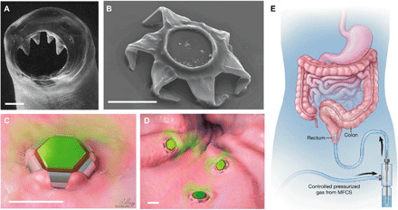
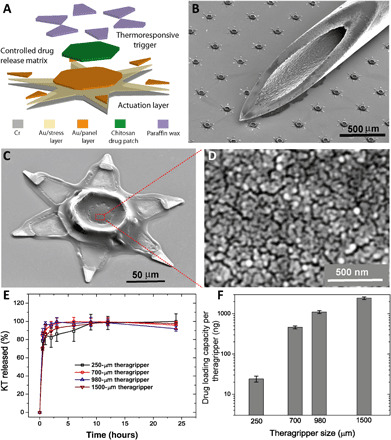
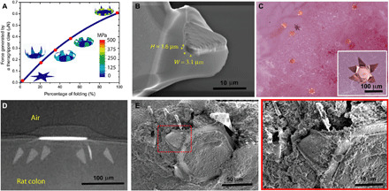
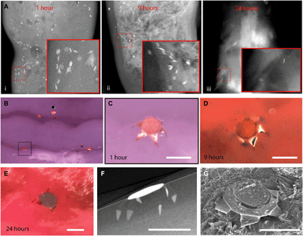
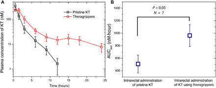
Extended-release gastrointestinal (GI) luminal delivery substantially increases the ease of administration of drugs and consequently the adherence to therapeutic regimens. However, because of clearance by intrinsic GI motility, device gastroretention and extended drug release over a prolonged duration are very challenging. Here, we report that GI parasite-inspired active mechanochemical therapeutic grippers, or theragrippers, can reside within the GI tract of live animals for 24 hours by autonomously latching onto the mucosal tissue. We also observe a notable sixfold increase in the elimination half-life using theragripper-mediated delivery of a model analgesic ketorolac tromethamine. These results provide first-in-class evidence that shape-changing and self-latching microdevices enhance the efficacy of extended drug delivery.


Figures

Fig. 1
Fig. 1. Shape-changing theragrippers as self-latching drug delivery devices.
(A) Scanning electron microscopy (SEM) image of the ventricular teeth of hookworm A. duodenale. The worm uses these sharp teeth to penetrate the mucosa and adheres in the GI tract for up to 2 years. Reprinted from Human Parasitology, 4th Ed. (18). Copyright 2013, with permission from Elsevier. (B) SEM image of a theragripper in the closed configuration. Like the hookworm, the theragrippers are equipped with sharp microtips. Schematic illustrations of (C) a single and (D) many theragrippers attached to the mucosal tissue and releasing encapsulated drug (colored in green). Scale bars, 100 μm (A to D). (E) Conceptual illustration of the rectal administration of drug-loaded theragrippers using a pressure-actuated microfluidic flow controller. Images (C) to (E) were illustrated by L. Gregg. MFCS, microfluidic flow control system.
Fig. 2
Fig. 2. Parallel fabrication of the theragrippers and their in vitro drug loading and release characteristics.
(A) Functional block diagram illustrating the microfabrication steps for an array of theragrippers, showing the actuation layer, drug-eluting layer, and the thermoresponsive trigger. (B) SEM image showing theragrippers next to the tip of a 22-gauge hypodermic needle. The theragrippers are small enough to pass safely through the GI tract without causing any gastric obstruction. (C) SEM image showing a single 250 μm, as fabricated theragripper with the drug-encapsulated chitosan patch at the center and the paraffin wax trigger layer on the hinges. (D) High-resolution SEM image showing the surface morphology of the chitosan patch at the center of the theragripper. The patch has pores less than 100 nm in size. (E) Release characteristics of ketorolac (KT) from theragrippers of four different sizes. (F) Plot showing the relative scaling of the drug loading capacity of theragrippers of different sizes. The entire loaded drug gets released over a period of 24 hours. While the 250-μm theragrippers were used for our in vivo experiments in rats, larger 1.5-mm theragrippers can be loaded with about 100 times more drug, for use in larger animal models and humans.
Fig. 3
Fig. 3. Theragrippers can apply sufficient force to penetrate the mucosa.
(A) Plot of the force generated by a theragripper as a function of the percentage of folding, generated by FEM. Each claw of the theragripper can generate a maximum force of around 0.6 μN per hinge. Insets show the simulated configurations at different stages of the folding process marked by red dots. The colors in the legend indicate the magnitude of the von Mises stress in the gripper. (B) Close-up SEM image of the tip of a theragripper, showing the cross section of the tip having a width (W) of approximately 3.1 μm and a height (H) of 1.6 μm. To estimate the pressure exerted by this tip as the gripper actuates, we used the Hertz contact mechanics model and assumed the tip to be a sphere of diameter 1.6 to 3.1 μm. (C) Ex vivo experiment showing many theragrippers latching onto the colon of a rat. The inset shows the bright-field zoomed-in image of a single theragripper. (D) μ-CT image of the cross section of a theragripper penetrating into the colon ex vivo. (E) SEM image of a theragripper latching onto the colon mucosa ex vivo. (F) Zoomed-in image of the red outlined region in (E), showing the penetration of the claw into the colon tissue.
Fig. 4
Fig. 4. Theragripper attachment to rat colon and retention upon rectal delivery.
(A) μ-CT images showing the retention of theragrippers in the rat colon (i) 1, (ii) 9, and (iii) 24 hours after rectal administration. The insets show zoomed-in images of the red outlined regions. (B) Postmortem optical examination of the rat colon showing attachment of several theragrippers, and (C) a zoomed-in image of the section shown by dotted lines in (B). (D and E) Optical image showing a theragripper attached to the colon (D) 9 and (E) 24 hours after rectal administration. (F) Postmortem μ-CT image showing the claws of a theragripper latching into the colon luminal surface in vivo. (G) SEM image showing the attachment of the theragripper to the mucosa after rectal administration. Scale bars, 100 μm. (C), (F), and (G) are obtained 1 hour after rectal administration of the theragrippers in the rat.
Fig. 5
Fig. 5. Theragrippers extend the delivery of ketorolac in vivo.
(A) Plot of the plasma concentration of ketorolac measured in rats as a function of time after pristine or free drug (black) and theragripper (red) were administered intrarectally. (B) AUClast comparison for pristine drug and theragripper-formulated drug delivery, showing that the drug exposure increases by approximately twofold with the theragrippers. P = 0.0283 for AUClast comparison in (B) is calculated using one-sided Student’s t test; N = 5 to 7.

References

    1. Traverso G., Langer R., Perspective: Special delivery for the gut. Nature 519, S19 (2015). - PubMed
    1. Garg R., Gupta G. D., Progress in controlled gastroretentive delivery systems. Trop. J. Pharm. Res. 7, 1055–1066 (2008).
    1. Jannin V., Lemagnen G., Gueroult P., Larrouture D., Tuleu C., Rectal route in the 21st Century to treat children. Adv. Drug Deliv. Rev. 73, 34–49 (2014). - PubMed
    1. Lowry M., Rectal drug administration in adults: How, when, why. Nurs. Times 112, 12–14 (2016). - PubMed
    1. Healthprize, “Medication adherence: Pharma’s $637 billion opportunity”; https://healthprize.com/blog/medication-adherence-pharmas-637-billion-op....

Publication types

Substances

LinkOut - more resources