. 2020 Oct 28;6(44):eabd2688.
doi: 10.1126/sciadv.abd2688. Print 2020 Oct.

Neutrophil-mediated carbamylation promotes articular damage in rheumatoid arthritis

Affiliations

Neutrophil-mediated carbamylation promotes articular damage in rheumatoid arthritis

Liam J O'Neil et al. Sci Adv. .

Abstract

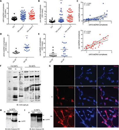
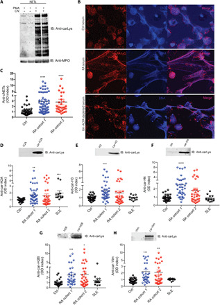
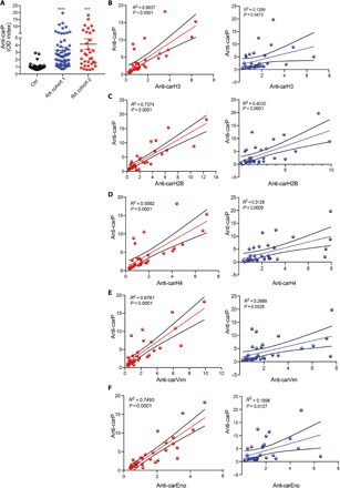
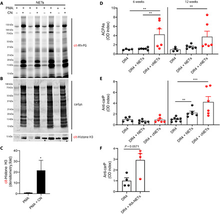
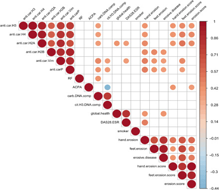
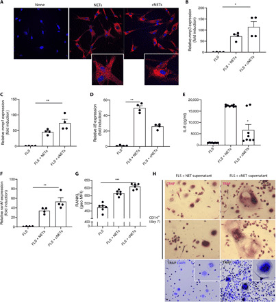
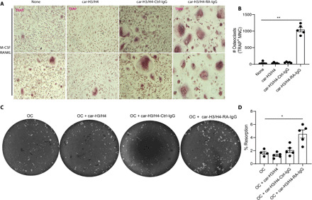
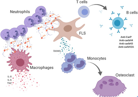
Formation of autoantibodies to carbamylated proteins (anti-CarP) is considered detrimental in the prognosis of erosive rheumatoid arthritis (RA). The source of carbamylated antigens and the mechanisms by which anti-CarP antibodies promote bone erosion in RA remain unknown. Here, we find that neutrophil extracellular traps (NETs) externalize carbamylated proteins and that RA subjects develop autoantibodies against carbamylated NET (cNET) antigens that, in turn, correlate with levels of anti-CarP. Transgenic mice expressing the human RA shared epitope (HLADRB1* 04:01) immunized with cNETs develop antibodies to citrullinated and carbamylated proteins. Furthermore, anti-carbamylated histone antibodies correlate with radiographic bone erosion in RA subjects. Moreover, anti-carbamylated histone-immunoglobulin G immune complexes promote osteoclast differentiation and potentiate osteoclast-mediated matrix resorption. These results demonstrate that carbamylated proteins present in NETs enhance pathogenic immune responses and bone destruction, which may explain the association between anti-CarP and erosive arthritis in RA.


Figures

Fig. 1
Fig. 1. Carbamylated proteins are increased in ionophore-generated NETs and in serum from RA subjects.
(A) Citrullinated histone H3–dsDNA complexes were measured in plasma from healthy controls (n = 25), RA cohort 1 (blue, n = 35), and RA cohort 2 (red, n = 34). Results are the mean ± SEM. Kruskal-Wallis test was used. *P < 0.05 and ****P < 0.0001. (B) Carbamylated lysine–dsDNA complexes were measured in plasma from healthy control (n = 25), RA cohort 1 (blue, n = 35), and RA cohort 2 (red, n = 34) (C) Citrullinated histone H3 and DNA complexes correlate with carbamylated lysine–dsDNA complexes. (D) Citrullinated histone H3–dsDNA complexes were measured in SF from OA (n = 10) and RA (n = 17). Results are the mean ± SEM. Mann-Whitney U test was used. (E) Carbamylated lysine–dsDNA complexes were measured in SF from OA (n = 10) and RA (n = 17) patients. Results are the mean ± SEM. Mann-Whitney U test was used. (F) Western blot analysis of carbamylated proteins in PMA, ionophore (Io)–generated NETs, and spontaneously generated NETs from RA patients. Representative picture of three independent experiments. (G) Immunofluorescence detection of carbamylated proteins in ionophore-generated NETs. Representative picture of three independent experiments. Carbamylated proteins are in red; DNA is blue. Original magnification, ×400. Ionophore-generated NETs were immunoprecipitated (IP) using anti–carbamylated lysine antibody. Immunoblot (IB) analysis was performed against (H) histone H3 or (I) histone H4. IgG was used as negative control. Lc, light chain.
Fig. 2
Fig. 2. Autoantibodies against cNET proteins are present in serum from RA patients.
(A) Western blot analysis shows increased cNET proteins after incubation with CN. RA IgGs recognize highly carbamylated (CN + PMA) NETs by (B) immunofluorescence and (C) ELISA analysis. Red, RA IgG; blue, DNA. Results are the mean ± SEM. Kruskal-Wallis test was used. Sera from healthy control (n = 25), RA cohort 1 (blue, n = 35), RA cohort 2 (red, n = 34), and SLE (n = 12) were analyzed for the presence of antibodies against carbamylated histones (D) H2A, (E) H3, (F) H4, and (G) H2B and against (H) vimentin. Results are the mean ± SEM. Kruskal-Wallis test was used. *P < 0.05, **P < 0.01, ***P < 0.001, and ****P < 0.0001. Western blot analysis was used to confirm carbamylation of each antigen [top panels of (D) to (H)]. Original magnification, ×400.
Fig. 3
Fig. 3. Autoantibodies against cNET antigens correlate with anti-CarP antibodies.
(A) Serum anti-CarP levels were measured in healthy control (n = 25), RA cohort 1 (blue, n = 35), and RA cohort 2 (red, n = 34). Results are the mean ± SEM. Kruskal-Wallis test was used. ***P < 0.001 and ****P < 0.0001. Correlation of anti-CarP antibodies with antibodies against carbamylated (B) histone H3, (C) histone H2B, (D) histone H4, (E) vimentin, and (F) α-enolase in two RA cohorts (red and blue).
Fig. 4
Fig. 4. DRB1*04:01 (DR4) transgenic mice that receive intra-articular injections of cNETs develop ACPAs and anti-CarP.
(A) Rh-PG probe against citrulline was used to detect specific citrullinated proteins in NETs (PMA) and cNETs (PMA + CN). (B) Western blot analysis against carbamylated proteins was performed in NET and cNETs. (C) Densitometry analysis of citrullinated histone H3 in NETs generated with PMA or PMA + CN (n = 4 per group). Results are the mean ± SEM. Mann-Whitney U test was used. *P < 0.05. (D) Serum ACPAs and (E and F) anti-CarP antibody levels were measured at various points in animals immunized with NETs, cNETs, or spontaneously generated RA-NETs (n = 4 to 8 per group). Results are the mean ± SEM. Kruskal-Wallis test was used. **P < 0.01 and ***P < 0.001.
Fig. 5
Fig. 5. Autoantibodies against carbamylated histones correlate with bone erosion.
Visual representation of significant correlations of autoantibodies against cNET proteins, carbamylated protein–DNA complexes, anti-CarP, ACPAs, and rheumatoid factor (RF) measured in RA subjects with clinical outcomes (n = 34) (global health, smoking, DAS28-ESR, hand and feet erosion score). Color gradient represents R2 values.
Fig. 6
Fig. 6. cNETs up-regulate RANKL in FLS and promote OC formation.
NETs and cNETs were incubated with RA FLS for 24 hours. (A) NETs and cNETs were internalized by RA FLS. Red, MPO; blue, DNA. Results are representative of three independent experiments. Original magnification, ×400. Quantitative polymerase chain reaction (qPCR) analysis shows (B) MMP3, (C) MMP1, (D) IL8, and (F) RANKL mRNA expression and ELISA analysis of (E) IL8 in RA FLS in the presence of NET and cNETs. Results are the mean ± SEM of four to eight independent experiments. Kruskal-Wallis test was used. *P < 0.05, **P < 0.01, and ***P < 0.001. (G) Plasma membrane RANKL was quantified by flow cytometry in RA FLS treated with NETs or cNETs for 24 hours. MFI, median fluorescent intensity. (H) CD14+ monocytes were incubated with supernatants of RA FLS treated with NETs or cNETs for 7 days. The presence of OCs was defined as multinucleated, TRAP (magenta, black for lower panel)–positive cells; nuclei are in blue (lower panel). Results are representative of three independent experiments. Original magnification, ×400 and ×200 (lower panels).
Fig. 7
Fig. 7. Anti–carbamylated histone H3 and H4–IgG complexes increase osteoclastogenesis and bone matrix resorption.
(A) Representative pictures of M-CSF/RANKL–mediated osteoclastogenesis in the presence or absence of anti–carbamylated histone H3/H4–IgG immune complexes. Original magnification, ×400. (B) Quantification of TRAP-positive multinucleated cells (MNC). Results are the mean ± SEM of four to five independent experiments. Kruskal-Wallis test was used. **P < 0.01. (C) Representative pictures of resorption pit assay of equal number of M-CSF/RANKL–generated OCs in the presence or absence of anti–carbamylated histone H3/H4-IgG immune complexes in a calcium phosphate plate. (D) Percentage of eroded surface using ImageJ. Results are the mean ± SEM of four to five independent experiments. Mann-Whitney U test was used. *P < 0.05.
Fig. 8
Fig. 8. Schematic representation of the role of carbamylation and anti-carbamylated immune responses in joint destruction in RA.
Infiltrating neutrophils release NETs containing carbamylated proteins. cNETs activate tissue macrophages to release proinflammatory cytokines. cNETs are internalized by FLS and up-regulate FLS production of RANKL that promotes osteoclastogenesis in CD14+ monocytes. Internalized cNETs by FLS can promote an adaptive immune response leading to generation of anti–carbamylated histone antibodies. In turn, these antibodies can form carbamylated immune complexes that promote OC formation and increase bone resorption.

Comment in

References

    1. Holers V. M., Autoimmunity to citrullinated proteins and the initiation of rheumatoid arthritis. Curr. Opin. Immunol. 25, 728–735 (2013). - PMC - PubMed
    1. Fisher B. A., Venables P. J., Inhibiting citrullination in rheumatoid arthritis: Taking fuel from the fire. Arthritis Res. Ther. 14, 108 (2012). - PMC - PubMed
    1. Brink M., Hansson M., Mathsson L., Jakobsson P.-J., Holmdahl R., Hallmans G., Stenlund H., Rönnelid J., Klareskog L., Rantapää-Dahlqvist S., Multiplex analyses of antibodies against citrullinated peptides in individuals prior to development of rheumatoid arthritis. Arthritis Rheum. 65, 899–910 (2013). - PubMed
    1. Arend W. P., Firestein G. S., Pre-rheumatoid arthritis: Predisposition and transition to clinical synovitis. Nat. Rev. Rheumatol. 8, 573–586 (2012). - PubMed
    1. Shi J., Knevel R., Suwannalai P., van der Linden M. P., Janssen G. M., van Veelen P. A., Levarht N. E., van der Helm-van Mil A. H., Cerami A., Huizinga T. W., Toes R. E., Trouw L. A., Autoantibodies recognizing carbamylated proteins are present in sera of patients with rheumatoid arthritis and predict joint damage. Proc. Natl. Acad. Sci. U.S.A. 108, 17372–17377 (2011). - PMC - PubMed

Publication types