. 2020 Oct 28;5(5):e00798-20.
doi: 10.1128/mSphere.00798-20.

Rift Valley Fever Virus Infection Causes Acute Encephalitis in the Ferret

Affiliations

Rift Valley Fever Virus Infection Causes Acute Encephalitis in the Ferret

Dominique J Barbeau et al. mSphere. .

Abstract

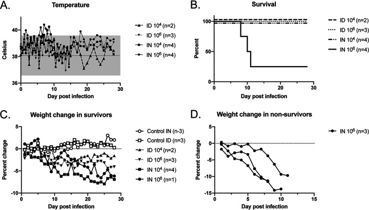
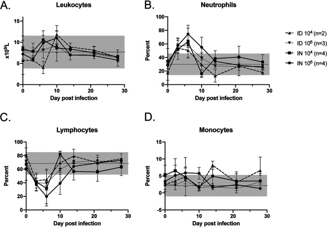
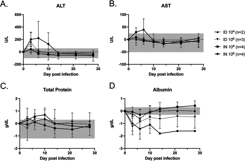
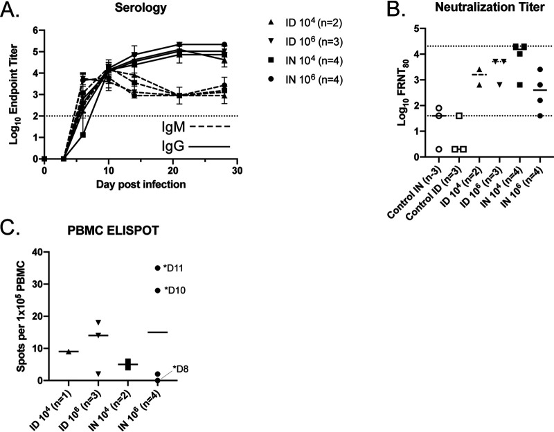
Rift Valley fever virus (RVFV) is a pathogen of both humans and livestock in Africa and the Middle East. Severe human disease is associated with hepatitis and/or encephalitis. Current pathogenesis studies rely on rodents and nonhuman primates, which have advantages and disadvantages. We evaluated disease progression in Mustela putorius furo (the ferret) following intradermal (i.d.) or intranasal (i.n.) infection. Infected ferrets developed hyperpyrexia, weight loss, lymphopenia, and hypoalbuminemia. Three of four ferrets inoculated intranasally with RVFV developed central nervous system (CNS) disease that manifested as seizure, ataxia, and/or hind limb weakness at 8 to 11 days postinfection (dpi). Animals with clinical CNS disease had transient viral RNAemia, high viral RNA loads in the brain, and histopathological evidence of encephalitis. The ferret model will facilitate our understanding of how RVFV accesses the CNS and has utility for the evaluation of vaccines and/or therapeutics in preventing RVFV CNS disease.IMPORTANCE Animal models of viral disease are very important for understanding how viruses make people sick and for testing out drugs and vaccines to see if they can prevent disease. In this study, we identify the ferret as a model of encephalitis caused by Rift Valley fever virus (RVFV). This novel model will allow researchers to evaluate ways to prevent RVFV encephalitis.

Keywords: Rift Valley fever virus; animal model; encephalitis; ferret; pathogenesis.


Figures

FIG 1
FIG 1
Clinical findings in RVFV-infected ferrets. Temperature measurements (A), survival (B), and weight changes (C and D) in ferrets inoculated with two different doses of rWT RVFV via the i.n. or i.d. route are shown. Numbers of animals in each group are noted. For panel A, the dotted line denotes the average of the values obtained in control animals over the course of the experiment, and the gray shading denotes 2 standard deviations from the averages.
FIG 2
FIG 2
Leukocyte changes in RVFV-infected ferrets. Total leukocytes (A) and the percentages of neutrophils (B), lymphocytes (C), and monocytes (D) are shown. Dotted lines denote the averages of the values obtained in control animals over the course of the experiment, and the gray shading in each graph denotes 2 standard deviations from the averages. Error bars indicate the standard deviations of the means.
FIG 3
FIG 3
Blood chemistry changes in RVFV-infected ferrets. Measurements of liver function (ALT and AST), total protein, and albumin, as indicated, are shown as a function of change from baseline. The gray shading in each graph denotes the range of changes observed from baseline in the control animals. Error bars indicate the standard deviations of the means.
FIG 4
FIG 4
Viral RNA loads in RVFV-infected ferrets. Viral RNA levels were measured in plasma over time (x axis, day postinfection) (A) and in various tissues at the time of euthanasia (B). The dotted line represents the limit of detection of the assays. Open symbols indicate isolation of virus.
FIG 5
FIG 5
Immunologic response in RVFV-infected ferrets. Average anti-RVFV IgM (dashed lines) and IgG (solid lines) enzyme-linked immunosorbent assay endpoint titers as measured over time (A) and neutralizing antibody titers at the time of euthanasia (B) are shown. The dotted lines represent the limits of detection of the assays. Error bars indicate the standard deviations of the means. (C) Virus-specific cellular immunity as measured by ELISPOT assay. Days (D) of euthanasia are indicated for the three animals that required euthanasia.
FIG 6
FIG 6
Pathologic findings in RVFV-infected ferrets. (A) Photograph of a grossly abnormal brain obtained during the necropsy of a high-dose i.n. infected ferret. The brain was characterized by diffuse hyperemia (top). An uninfected control brain is shown for comparison (bottom). (B) Foci (boxed) of hepatic inflammation and necrosis were noted in the liver of an animal infected with RVFV i.n. at 104 TCID50 (magnification, ×20). (C) Viral RNA detection in the lungs of one animal infected with RVFV i.n. at 106 TCID50 corresponded with diffuse pneumonitis and type II pneumocyte proliferation (arrow) in the setting of lung collapse (magnification, ×40). Perivascular infiltrates (boxed, ×20 magnification) (D), meningitis (arrow, ×20 magnification) (E), choroiditis (boxed, ×10 magnification) (F), glial nodules (boxed, ×20 magnification) (G), and laminar necrosis (arrows, ×10 magnification) (H) were noted in the brains of animals with encephalitis. Representative CNS photos are shown from animals infected with RVFV i.n. at 106 TCID50. Scale bar, 100 μm.

References

    1. Jost CC, Nzietchueng S, Kihu S, Bett B, Njogu G, Swai ES, Mariner JC. 2010. Epidemiological assessment of the Rift Valley fever outbreak in Kenya and Tanzania in 2006 and 2007. Am J Trop Med Hyg 83:65–72. doi:10.4269/ajtmh.2010.09-0290. - DOI - PMC - PubMed
    1. LaBeaud AD, Muiruri S, Sutherland LJ, Dahir S, Gildengorin G, Morrill J, Muchiri EM, Peters CJ, King CH. 2011. Postepidemic analysis of Rift Valley fever virus transmission in northeastern Kenya: a village cohort study. PLoS Negl Trop Dis 5:e1265. doi:10.1371/journal.pntd.0001265. - DOI - PMC - PubMed
    1. Meegan JM, Hoogstraal H, Moussa MI. 1979. An epizootic of Rift Valley fever in Egypt in 1977. Vet Rec 105:124–125. doi:10.1136/vr.105.6.124. - DOI - PubMed
    1. Swai ES, Schoonman L. 2009. Prevalence of Rift Valley fever immunoglobulin G antibody in various occupational groups before the 2007 outbreak in Tanzania. Vector Borne Zoonotic Dis 9:579–582. doi:10.1089/vbz.2008.0108. - DOI - PubMed
    1. Madani TA, Al-Mazrou YY, Al-Jeffri MH, Mishkhas AA, Al-Rabeah AM, Turkistani AM, Al-Sayed MO, Abodahish AA, Khan AS, Ksiazek TG, Shobokshi O. 2003. Rift Valley fever epidemic in Saudi Arabia: epidemiological, clinical, and laboratory characteristics. Clin Infect Dis 37:1084–1092. doi:10.1086/378747. - DOI - PubMed

Publication types

LinkOut - more resources