

Bilateral Central Retinal Vein Occlusion in a 40-Year-Old Man with Severe Coronavirus Disease 2019 (COVID-19) Pneumonia
- PMID: 33116072
- PMCID: PMC7603800
- DOI: 10.12659/AJCR.927691
Bilateral Central Retinal Vein Occlusion in a 40-Year-Old Man with Severe Coronavirus Disease 2019 (COVID-19) Pneumonia
Abstract
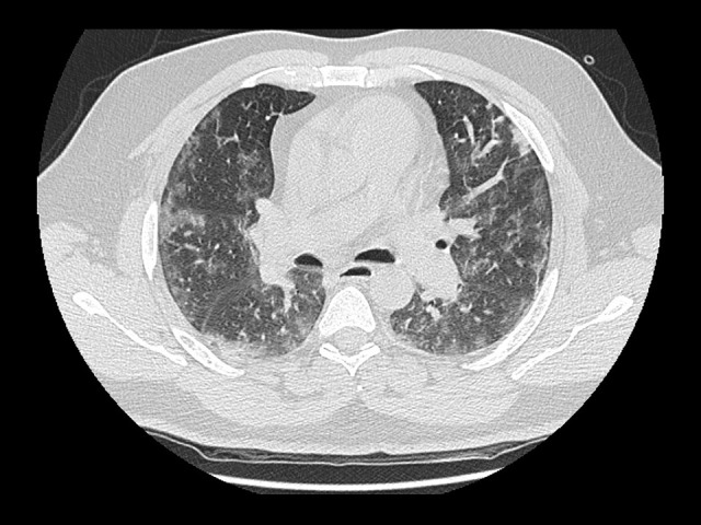
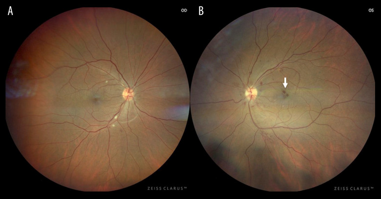
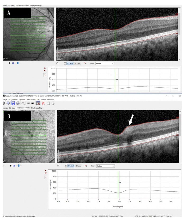
BACKGROUND COVID-19 is the disease caused by the novel virus, severe acute respiratory syndrome Coronavirus 2 (SARS-CoV-2). The spectrum of disease seen in patients with COVID-19 infection ranges from asymptomatic or mild symptoms to severe pneumonia and even acute respiratory distress syndrome, which often requires invasive ventilation and intensive care. COVID-19-associated infection can be catastrophic, leading to both arterial and venous occlusion, microinfarcts, and multiorgan failure, although retinal vein occlusion has not yet been reported. CASE REPORT We present the case of a 40-year-old man who presented with a 3-day history of shortness of breath, cough, and fever. He also reported right calf pain and blurring of vision in both eyes. His medical history included hypertension and morbid obesity. The patient was found to have severe COVID-19 pneumonia on high-resolution computed tomography of the chest, right leg deep venous thrombosis on Doppler ultrasonography, and bilateral central retinal vein occlusion (RVO) on fundal examination. He was started on full-dose anticoagulation and discharged on rivaroxaban for 3 months. After 2 weeks of therapy, he had fully recovered from his COVID-19 symptoms and had near-normal vision. CONCLUSIONS COVID-19 infection can cause RVO. Early full-dose anticoagulation should be considered in high-risk patients with severe COVID-19 infection. Ophthalmologists and other clinicians should have a high index of suspicion for RVO in patients with COVID-19 infection who presenting with blurred vision and severe pneumonia.
Conflict of interest statement
None
Figures
References
-
- World Health Organization Coronavirus Disease Dashboard (COVID-19) https://covid19.who.int/
-
- World Health Organisation (WHO) Use of laboratory methods for SARS diagnosis. 2020. https://who.int/csr/sars/labmethods/en/
-
- Coscas G, Loewenstein A, Augustin A, et al. Management of retinal vein occlusion – consensus document. Ophthalmologica. 2011;226:4–28. - PubMed
Publication types
MeSH terms
LinkOut - more resources
Full Text Sources
Miscellaneous
