. 2020 Oct 28;11(1):5432.
doi: 10.1038/s41467-020-19230-w.

Identification of a myotropic AAV by massively parallel in vivo evaluation of barcoded capsid variants

Affiliations

Identification of a myotropic AAV by massively parallel in vivo evaluation of barcoded capsid variants

Jonas Weinmann et al. Nat Commun. .

Abstract

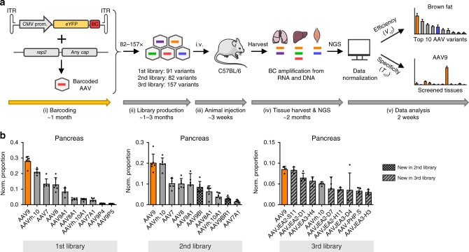
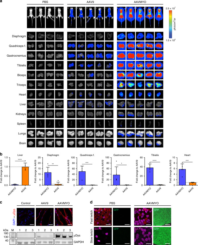
Adeno-associated virus (AAV) forms the basis for several commercial gene therapy products and for countless gene transfer vectors derived from natural or synthetic viral isolates that are under intense preclinical evaluation. Here, we report a versatile pipeline that enables the direct side-by-side comparison of pre-selected AAV capsids in high-throughput and in the same animal, by combining DNA/RNA barcoding with multiplexed next-generation sequencing. For validation, we create three independent libraries comprising 183 different AAV variants including widely used benchmarks and screened them in all major tissues in adult mice. Thereby, we discover a peptide-displaying AAV9 mutant called AAVMYO that exhibits superior efficiency and specificity in the musculature including skeletal muscle, heart and diaphragm following peripheral delivery, and that holds great potential for muscle gene therapy. Our comprehensive methodology is compatible with any capsids, targets and species, and will thus facilitate and accelerate the stratification of optimal AAV vectors for human gene therapy.


Conflict of interest statement

D.G. is a co-founder and shareholder of AaviGen GmbH. Part of this work was conducted as a sponsored research agreement between the Grimm lab and Boehringer Ingelheim Pharma GmbH & Co. KG. J.W. has performed the studies reported here during his doctoral work at Heidelberg University Hospital and is now an employee of Boehringer Ingelheim Pharma GmbH & Co. KG. D.G., J.E.A. and J.W. are inventors on a pending patent application (International application number: PCT/EP2019/060790; Publication number: WO/2019/207132) covering AAVMYO. T.S., M.L., and T.L. are employees of Boehringer Ingelheim Pharma GmbH & Co. KG. All other authors declare no competing interests.

Figures

Fig. 1
Fig. 1. A robust workflow for massively parallel in vivo AAV capsid stratification.
a Experimental setup. Shown are the essential experimental and bioinformatic steps, from the (i) cloning of barcoded YFP reporter vectors, (ii) library production, and (iii) animal (mouse here) injection, to (iv) tissue harvest and NGS, followed by (v) data analysis. Also shown is a timeline for the entire workflow or the individual steps. Note that the exact time required in step (ii) depends on the size of the library. Likewise, the time to perform the final bioinformatic analysis in step (v) is determined by available computing (IT) power. The first step (orange arrow) can be skipped if users start with our pre-existing collection of barcoded vector genomes, which will cut down the required overall time to <6 months. Please see the main text for further details. b Ranking of capsids in all three libraries by transcriptional efficiency (Vαβ) in the pancreas. Shown are the top ten AAV variants and the proportion of their transcriptional efficiency after normalization to all capsids or barcodes, respectively, in each library. Depicted values are the average from six C57BL/6 J mice with SD. BC, barcode; i.v., intravenous. This figure contains clipart from Servier Medical Art. Source data are available in the Source data file.
Fig. 2
Fig. 2. Examples for robust capsids in the liver, lung, brain, and musculature.
a Top 10 AAV variants in the second library and the shown tissues based on normalized transcriptional efficiency (Vαβ). Depicted values are the average from six C57BL/6 J mice with SD. b Transcriptional specificity (Tαβ) of the shown four capsids as normalized proportion per cell (diploid genome, dg) in abdominal aorta (Aa), thoracic aorta (At), brain (B), blood cells (BlC), colon (C), diaphragm (Di), duodenum (Du), eye, brown fat (FatB), white fat (FatW), heart (H), inner ear (I), kidney (K), liver (Li), lung (Lu), ovaries (O), pancreas (P), quadriceps femoris (QF), spleen (S), and stomach (St). Depicted are average cDNA values from six C57BL/6 J mice with SD. Colors in a and b highlight the same capsids in both panels. Source data are available in the Source data file.
Fig. 3
Fig. 3. Validation of the myotropic AAV capsid AAVMYO.
a Transcriptional specificity (Tαβ) of the shown capsids from the third library as normalized proportion per cell in aorta (A), biceps (Bi), colon (C), CD11b-, CD11c-, CD19-, or CD3-positive cells, diaphragm (Di), duodenum (Du), eye, brown fat (FatB), white fat (FatW), heart (H), inner ear (I), kidney (K), liver (Li), lung (Lu), ovaries (O), pancreas (P), quadriceps femoris (QF), and stomach (St). Depicted are average cDNA values from six C57BL/6 J mice with SD. b Comparison of AAVMYO to AAV9 and AAVpo.1 after individual i.v. injection. Shown are relative eyfp mRNA quantities in the liver, diaphragm, quadriceps femoris, and heart. AAV9 values were always set to 1 and the others depicted as fold changes. Relative quantities (2−ΔΔCt) of viral eyfp transcripts were measured via RT-qPCR and a POLR2A housekeeper. Depicted values are the average of three C57BL/6 J mice with SD. Colors in a and b highlight the same capsids in both panels; *p < 0.05, **p < 0.01, ***p < 0.001, and ****p < 0.0001 (one-way ANOVA with Tukey’s multiple comparison test). c Representative images of dissected C57BL/6 J mice (left: ventral; right: dorsal position) that were i.v. injected with 5 × 1011 vg per mouse and sacrificed two weeks later. d Representative 10 µm cryosections (n = 8 replicates for AAV9, AAVMYO and n = 3 for PBS) of the liver, diaphragm, heart, biceps, and quadriceps femoris of the mice from c. Direct EGFP fluorescence was detected (green) together with the DAPI signal (blue). Scale bars are 1 mm (full sections) or 100 µm (10× magnifications). Exposure settings were normalized to the liver of the AAV9 group. Source data are available in the Source data file.
Fig. 4
Fig. 4. Additional validation of AAVMYO.
a Shown on top are whole-body images of luciferase expression in CB17-SCID mice 4 weeks after i.v. injection with the shown vectors. Shown underneath are images of luciferase expression in the indicated organs from the same mice, harvested 1 week later. b Shown are fold-changes of luciferase mRNA expression of each organ as determined by qRT-PCR in the AAVMYO cohort versus the AAV9 group. Depicted values are the average of the five CB17-SCID mice from panel a with SD; *p < 0.05, **p < 0.01, ***p < 0.001, and ****p < 0.0001 (unpaired two-tailed t test). c Comparison of µDys expression in quadriceps femoris sections (n = 4 replicates for all groups) and protein lysates of mdx mice 4 weeks after i.v. injection of 2 × 1011 vg (for IHC) or 1 × 1012 vg (for western blot) of a control vector (AAVMYO expressing Firefly luciferase), or of AAV9 or AAVMYO encoding µDys. d Staining of muscle fibers type I (BA-F8), type IIa (sc-71), type IIb (BF-F3), and GFP in quadriceps femoris sections of C57BL/6 mice (n = 4 replicates for all groups) injected with 5 × 1011 vg of AAVMYO. Scale bars for c and d are 25 and 50 µm, respectively. Source data are available in the Source data file.

References

    1. Grimm D, Zolotukhin SE. Pluribus unum: 50 years of research, millions of viruses, and one goal-tailored acceleration of AAV evolution. Mol. Ther. 2015;23:1819–1831. doi: 10.1038/mt.2015.173. - DOI - PMC - PubMed
    1. Herrmann AK, et al. A robust and all-inclusive pipeline for shuffling of adeno-associated viruses. ACS Synth. Biol. 2019;8:194–206. doi: 10.1021/acssynbio.8b00373. - DOI - PubMed
    1. Deverman BE, et al. Cre-dependent selection yields AAV variants for widespread gene transfer to the adult brain. Nat. Biotechnol. 2016;34:204–209. doi: 10.1038/nbt.3440. - DOI - PMC - PubMed
    1. Korbelin J, et al. Pulmonary targeting of adeno-associated viral vectors by next-generation sequencing-guided screening of random capsid displayed peptide libraries. Mol. Ther. 2016;24:1050–1061. doi: 10.1038/mt.2016.62. - DOI - PMC - PubMed
    1. Yang L, et al. A myocardium tropic adeno-associated virus (AAV) evolved by DNA shuffling and in vivo selection. Proc. Natl Acad. Sci. USA. 2009;106:3946–3951. doi: 10.1073/pnas.0813207106. - DOI - PMC - PubMed

Publication types

Substances

LinkOut - more resources