. 2020 Oct 28;11(1):5453.
doi: 10.1038/s41467-020-19145-6.

ACE2 localizes to the respiratory cilia and is not increased by ACE inhibitors or ARBs

Affiliations

ACE2 localizes to the respiratory cilia and is not increased by ACE inhibitors or ARBs

Ivan T Lee et al. Nat Commun. .

Abstract

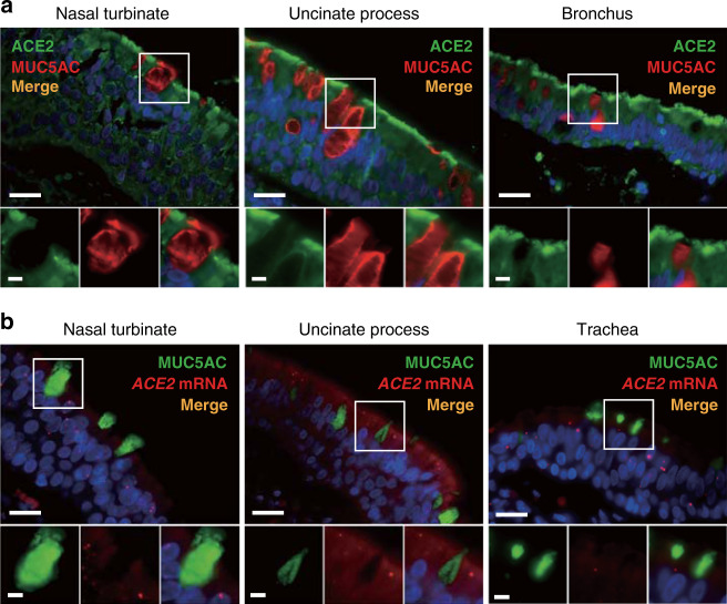
The coronavirus SARS-CoV-2 is the causative agent of the ongoing severe acute respiratory disease pandemic COVID-19. Tissue and cellular tropism is one key to understanding the pathogenesis of SARS-CoV-2. We investigate the expression and subcellular localization of the SARS-CoV-2 receptor, angiotensin-converting enzyme 2 (ACE2), within the upper (nasal) and lower (pulmonary) respiratory tracts of human donors using a diverse panel of banked tissues. Here, we report our discovery that the ACE2 receptor protein robustly localizes within the motile cilia of airway epithelial cells, which likely represents the initial or early subcellular site of SARS-CoV-2 viral entry during host respiratory transmission. We further determine whether ciliary ACE2 expression in the upper airway is influenced by patient demographics, clinical characteristics, comorbidities, or medication use, and show the first mechanistic evidence that the use of angiotensin-converting enzyme inhibitors (ACEI) or angiotensin II receptor blockers (ARBs) does not increase susceptibility to SARS-CoV-2 infection through enhancing the expression of ciliary ACE2 receptor. These findings are crucial to our understanding of the transmission of SARS-CoV-2 for prevention and control of this virulent pathogen.


Conflict of interest statement

The authors declare no competing interests.

Figures

Fig. 1
Fig. 1. Immunohistochemical analysis of ACE2 protein localization across human tissues using multiple anti-ACE2 antibodies.
Representative images of human tissues on a tissue microarray (TMA) stained by chromogenic immunohistochemistry using antibodies targeting the ACE2 protein (brown) and counterstained with hematoxylin (blue). Highest ACE2 expression was observed in the villi of the intestinal tract (jejunum), renal tubules, testis, and glandular cells in the seminal vesicle. Minimal to no/non-specific staining can be seen in the heart, stomach, spleen, skin, and liver. Staining of lung pneumocytes was observed using Abcam ab15348, and less specifically with Sigma HPA000288 (Fig. 2b; Supplementary Table 1). Scale bars: 100 μm.
Fig. 2
Fig. 2. ACE2 protein expression within human vascular endothelial cells and the lung.
a Representative double immunofluorescence staining of ACE2 and endothelial cell marker CD31 in the blood vessels of human nasal turbinates using six different anti-ACE2 antibodies and anti-CD31. b Double immunofluorescence staining of ACE2 and type II pneumocyte marker mucin 1 (MUC1) in the human lung using six different anti-ACE2 antibodies and anti-MUC1. Abcam ab15348 clone yielded the most robust staining of pneumocytes, while the other clones showed negligible or less specific membrane staining. Scale bars: 20 μm (top) and 5 μm (bottom).
Fig. 3
Fig. 3. ACE2 protein expression in the cilia organelle of upper and lower respiratory tract epithelia and in a ciliated kidney epithelial cell line.
a Representative double immunofluorescence staining of ACE2 and acetylated α-tubulin (ACTUB) on normal human nasal turbinate, ethmoid sinus, uncinate process (sinus), trachea, and bronchus, using anti-ACE2 and anti-ACTUB antibodies, respectively. b Representative double immunofluorescence staining of ACE2 and ACTUB on normal C57BL/6J mouse nasal turbinate and trachea. c Immunofluorescent staining of (top panel) ACE2, cilia marker ADP-ribosylation factor-like protein 13B (ARL13B), and cilia centrosome marker FGFR1 oncogene partner (FOP); (bottom panel) ACE2, and cilia markers ACTUB and ARL13B in a ciliated mouse cell line, IMCD3. d Immunofluorescent staining of ACE2 in the primary cilia of IMCD3 cells transiently transfected with human ACE2 (yellow outline) compared to endogenous mouse ACE2 (blue outline). e Quantified percentages of endogenous ACE2-positive cilia (34.67 ± 13.58%; control (Ctrl)) versus cilia with overexpressed human ACE2 (82.67 ± 4.73%). Ciliated cells were identified by staining of ARL13B. Error bars represent mean ± SD. (n = 100 cells examined per experiment over three independent experiments). (Two-tailed Student’s t test, **p = 0.004). f Representative multiplexed images of in situ hybridization against the SARS-CoV-2 Spike mRNA, in combination with immunofluorescence staining of ACE2 and the differentiated epithelial cell marker cytokeratin 8 (KRT8). SARS-CoV-2 Spike mRNA expression (red) was detected within ciliated epithelial cells containing motile cilia positive for ACE2 (green). The nuclei were stained using DAPI (blue) as a counterstain. Scale bars: 20 μm (a, b top panels; f large panels); 5 μm (a, b bottom panels; f small panels); 2 μm (c, d).
Fig. 4
Fig. 4. ACE2 protein and mRNA expression are not found in secretory goblet cells of the human airway.
a Representative immunofluorescence double staining of ACE2 and mucin 5AC (MUC5AC) reveals absence of co-localization of ACE2 within secretory goblet cells in the human nasal turbinate, uncinate process, and bronchus. b Representative in situ hybridization using an ACE2 probe in combination with an anti-MUC5AC antibody. ACE2 mRNA expression (red dots) was not detected within goblet cells marked by MUC5AC in the nasal turbinate, uncinate process, and trachea. Nuclei were stained using DAPI. Scale bars: 20 μm (top) and 5 μm (bottom).
Fig. 5
Fig. 5. Comparison of ciliary ACE2 protein expression by age, sex, smoking status, sinus disease, and anatomical region.
a No statistically significant changes in ACE2 expression was detected among patients less than or greater than 65 years of age, males versus females, and patients with varying smoking history. (Two-tailed Mann–Whitney test or Kruskal–Wallis test, p > 0.05). b No statistically significant difference in ACE2 expression was observed between healthy controls and patients with chronic rhinosinusitis with polyps (CRSwNP) or without polyps (CRSsNP). (Kruskal–Wallis test, p > 0.05). c No statistically significant difference in ACE2 expression was noted between distinct human nasal tissue sites/regions. (Two-tailed Mann–Whitney test or Kruskal–Wallis test, p > 0.05). UNC uncinate process, Turb nasal turbinates, Eth ethmoid sinus, NP benign nasal polyps. The bottom and top of the box plots represent the 25th and 75th percentiles, respectively. The bands within the box show the median value, and the whiskers extending from both ends of the boxes are minimum and maximum values. Each dot represents one patient.
Fig. 6
Fig. 6. ACE2 expression in the nasal cilia is not increased in patients taking ACEI or ARBs.
a Quantification of ACE2 in controls and patients taking ARBs and ACEI. In the Stanford cohort, ACE2 is slightly but statistically significantly lower in patients taking ACEI (0.19 ± 0.02) compared to controls (0.26 ± 0.06). (Kruskal–Wallis test p = 0.021; Dunn’s multiple comparison post-hoc test, *adjusted p = 0.043). There were no statistically significant differences in ACE2 expression between patients taking ARBs and controls in the Stanford, National Taiwan University Hospital (NTUH), and China Medical University Hospital (CMUH) cohorts. b In the Stanford cohort, when including only controls with hypertension (HTN) on other medications (“HTN w/o ARBs/ACEI”), ACE2 expression was statistically different between the groups (Kruskal–Wallis test, p = 0.044) but Dunn’s multiple comparison post-hoc test did not reveal any statistical significance between the three groups. No statistically significant differences were seen among patients taking ARBs compared to controls. c When cohorts from all three institutions were normalized by Z-score and integrated, patients taking ACEI (−0.72 ± 0.42) had a lower ACE2 expression compared to controls with hypertension (0.41 ± 1.07). (Kruskal–Wallis test, p = 0.032; Dunn’s multiple comparison post-hoc test, *adjusted p = 0.043). Patients taking ARBs (−0.15 ± 0.95) showed a trend towards lower ACE2 compared to controls with hypertension, but this was not statistically significant. d ACE2 expression among patients of older (≥65 years) and younger (<65 years) age taking ARBs or ACEI was not statistically divergent from control patients of the same age group. (Kruskal–Wallis test, p > 0.05). e ACE2 expression among male and female patients on ARBs or ACEI trended comparably or lower than same-sex controls except for males taking ARBs in the CMUH group who showed a trend towards higher ACE2 expression. No statistically significant differences were observed. (Kruskal–Wallis test, p > 0.05). f Among non-smokers, there was a statistically significant trend towards lower ACE2 expression in patients taking ACEI compared to controls in the Stanford group (Kruskal–Wallis test, p = 0.021; Dunn’s multiple comparison post-hoc test, *adjusted p = 0.035). No statistical significance was observed with the non-smokers on ARBs. All data are noted as mean ± SD. Kruskal–Wallis test was used for three group comparisons and two-tailed Mann–Whitney test was used for two-group comparisons. Box plots are similar in format to Fig. 5.
Fig. 7
Fig. 7. Illustration of ACE2 expression in the motile cilia of epithelial cells in the nose, paranasal sinuses, trachea, and lower airways.
The luminal differentiated airway epithelial cells consist of ciliated columnar cells (~80%) and secretory goblet cells (~20%). Club cells are infrequently found in the human upper airway. The basal cell layer, which faces the lamina propria, is comprised of both basal and suprabasal cells, which are considered multipotent progenitors capable of renewing the airway epithelium. This schematic depicts how SARS-CoV-2 may bind to ACE2 expressed on the cilia of the nasal cavity following exposure to respiratory droplets or airborne particles.

Update of

References

    1. Wu F, et al. A new coronavirus associated with human respiratory disease in China. Nature. 2020;579:265–269. - PMC - PubMed
    1. Wang D, et al. Clinical characteristics of 138 hospitalized patients with 2019 novel coronavirus–infected pneumonia in Wuhan, China. JAMA. 2020;323:1061–1069. doi: 10.1001/jama.2020.1585. - DOI - PMC - PubMed
    1. Grasselli, G. et al. Baseline characteristics and outcomes of 1591 patients infected with SARS-CoV-2 admitted to ICUs of the Lombardy Region, Italy. JAMA323, 1574–1581 (2020). - PMC - PubMed
    1. Xie J, Tong Z, Guan X, Du B, Qiu H. Clinical characteristics of patients who died of coronavirus disease 2019 in China. JAMA Netw. Open. 2020;3:e205619. doi: 10.1001/jamanetworkopen.2020.5619. - DOI - PMC - PubMed
    1. CDC COVID-19 Response Team. et al. Preliminary estimates of the prevalence of selected underlying health conditions among patients with coronavirus disease 2019 - United States, February 12-March 28, 2020. MMWR Morb. Mortal. Wkly Rep. 2020;69:382–386. doi: 10.15585/mmwr.mm6913e2. - DOI - PMC - PubMed

Publication types

MeSH terms

Substances