. 2020 Oct 28;10(1):18529.
doi: 10.1038/s41598-020-75152-z.

Non-contact vital-sign monitoring of patients undergoing haemodialysis treatment

Affiliations

Non-contact vital-sign monitoring of patients undergoing haemodialysis treatment

Mauricio Villarroel et al. Sci Rep. .

Abstract

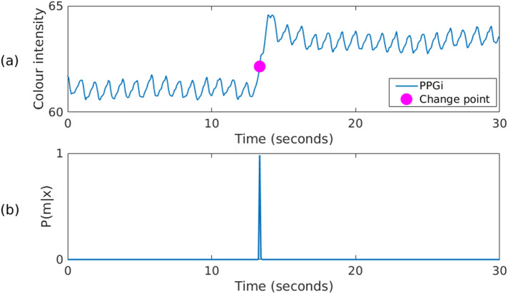
A clinical study was designed to record a wide range of physiological values from patients undergoing haemodialysis treatment in the Renal Unit of the Churchill Hospital in Oxford. Video was recorded for a total of 84 dialysis sessions from 40 patients during the course of 1 year, comprising an overall video recording time of approximately 304.1 h. Reference values were provided by two devices in regular clinical use. The mean absolute error between the heart rate estimates from the camera and the average from two reference pulse oximeters (positioned at the finger and earlobe) was 2.8 beats/min for over 65% of the time the patient was stable. The mean absolute error between the respiratory rate estimates from the camera and the reference values (computed from the Electrocardiogram and a thoracic expansion sensor-chest belt) was 2.1 breaths/min for over 69% of the time for which the reference signals were valid. To increase the robustness of the algorithms, novel methods were devised for cancelling out aliased frequency components caused by the artificial light sources in the hospital, using auto-regressive modelling and pole cancellation. Maps of the spatial distribution of heart rate and respiratory rate information were developed from the coefficients of the auto-regressive models. Most of the periods for which the camera could not produce a reliable heart rate estimate lasted under 3 min, thus opening the possibility to monitor heart rate continuously in a clinical environment.


Conflict of interest statement

The authors declare no competing interests.

Figures

Figure 1
Figure 1
Distribution of physiological values for all the patients in the clinical study. (a) Heart rate and (b) SpO2 from the finger pulse oximeter. (c) Respiratory rate from the chest belt.
Figure 2
Figure 2
Agreement between the reference heart rate values (computed from the agreement between the two pulse oximeters) and the camera estimates (computed using the ARk algorithm and the Kalman ND data fusion method) for the valid dialysis video data, comprising a total recording time of approximately 219.8 h. (a) The Bland–Altman plot presents no sensor bias. (b) The differences between the camera estimates and the reference heart rate values are normally distributed. (c) The distribution of the mean values shows that most of the heart rate estimates are within the expected physiological range for adults. (d) The plot shows a strong correlation between the two devices, with a correlation coefficient of 0.98.
Figure 3
Figure 3
Agreement between the reference respiratory rate values (computed from the ECG and chest belt) and the camera estimates computed over the regions of interest on the patients’ face for the valid dialysis video data, comprising a total recording time of approximately 101.4 h. (a) The Bland–Altman plot presents minimal sensor bias. (b) The differences between the two estimates. (c) The distribution of the mean values show that most of the respiratory rate estimates are within the expected physiological range for adults. (d) The plot shows a strong correlation between the two devices, with a correlation coefficient of 0.92.
Figure 4
Figure 4
Gaps in heart rate estimation using the Kalman ND data fusion method. Most of the periods for which the camera could not produce a heart rate estimate were under 3 min.
Figure 5
Figure 5
A typical dialysis data collection set-up. The red circle shows the location of the video camera. A pulse oximeter is attached to the patient’s finger on the right hand. The Equivital EQ-02 LifeMonitor is located on the patient’s chest. Consent was obtained from the patient to use the image.
Figure 6
Figure 6
The process of extracting PPGi signals. (a) Activity Index computed for a typical 4-h dialysis session with screenshots above showing the patient: adjusting headphones to listen to music near minute 10, sleeping at minute 60, awake and engaging in a phone conversation at minute 120, sleeping at minute 200, and interacting with the clinical staff at minute 240. (b) The image at minute 60 showing: the patient’s detected face (green rectangle), ROIR (blue square), and a sample ROIS (red square). (c) Image segmentation output. (d) Location and size of the grid for all ROIS. (e) A sample 15-s PPGi time series extracted from the ROIS in (b). (f) Sample ROIR time series extracted from the ROIR in (b). Consent was obtained from the patient to use these images.
Figure 7
Figure 7
Bayesian change point detection algorithm applied to a 30-s window during which a fluorescent light was turned on, producing a step change in the recorded pixel intensity signal. (a) Input PPGi signal with the change point marked around second 13. (b) The probability of the change point.
Figure 8
Figure 8
Steps to compute the location of the onset of each cardiac pulse for a 10-s PPGi time series: (a) Input PPGi waveform. (b) Slope sum function (SSF). (c) Boxed Slope Sum Function (BSSF), scaled to match each pulse in the SSF function. (d) The adaptive threshold applied to the uBSSF signal. (e) The detected beat onsets.
Figure 9
Figure 9
Time alignment for multi-scale DTW. (a) Normalised input beat (blue) and template (red). (b) The accumulated distance matrix with darker regions present areas where the beat and template are close, while brighter regions constitute areas where they are further apart; the optimal minimum path is shown in white. (c) DTW time alignment between the beat and template. (d) Finding the minimum path in the iterative multi-scale DTW algorithm.
Figure 10
Figure 10
Example of a poor-quality beat. The resultant combined SQIbeat value was 0. (a) Input template and beat, (b) rotated and normalised template and beat, (c) piece-wise linear approximation, (d) check if the beat was within physiological bounds for heart rate, (e) check if the beat was within the amplitude thresholds for the current window, (f) clipping detection, (g) DTW showing the minimum path with a cost of 28.10, (h) accumulated cost matrix showing the minimum path.
Figure 11
Figure 11
Example of a good-quality beat. The resultant combined SQIbeat value was 1. (a) Input template and beat, (b) rotated and normalised template and beat, (c) piece-wise linear approximation, (d) check if the beat was within physiological bounds for heart rate, (e) check if the beat was within the amplitude thresholds for the current window, (f) clipping detection, (g) DTW showing the minimum path with a cost of 3.58, (h) accumulated cost matrix showing the minimum path.
Figure 12
Figure 12
AR pole cancellation process for a 15-s window from the (a) ROIS and (b) ROIR. (c) Zero-pole plot and (d) frequency components for ROIS. (e) Zero-pole plot and (f) frequency components for ROIR. (g) Similar poles from ROIR in ROIS are cancelled creating a new autoregressive model ARk and (h) the power spectrum is computed from the remaining pole. The heart rate estimate is taken as the peak of the reconstructed power spectrum at 1.2 Hz, corresponding to a heart rate of approximately 72 beats/min. (i) From the remaining pole, a signal can be generated from a white noise input source.
Figure 13
Figure 13
ROI selection for respiratory rate estimation for (a) one sample image frame. Several ROI were defined for two main areas: (b) skin areas across the patient’s face defined on colour images, (c) the patient’s upper torso defined on grey-scale images. 30-s pixel intensity time series extracted from (d) the green channel from a ROI on the patient’s face, (e) grey-scale images from a ROI on the patient’s upper chest.

References

    1. Takano C, Ohta Y. Heart rate measurement based on a time-lapse image. Med. Eng. Phys. 2007;29:853–857. - PubMed
    1. Lee, K.-Z., Hung, P.-C. & Tsai, L.-W. Contact-free heart rate measurement using a camera. IEEE Can. Conf. Comput. Robot Vis. 147–152 (2012).
    1. Kranjec J, Beguš S, Geršak G, Drnovšek J. Non-contact heart rate and heart rate variability measurements: a review. Biomed. Signal Process. Control. 2014;13:102–112.
    1. AL-Khalidi F. Q, Saatchi R, Burke D, Elphick H, Tan S. Respiration rate monitoring methods: a review. Pediatr. Pulmonol. 2011;46:523–529. - PubMed
    1. Meredith, D. Continuous Monitoring During Haemodialysis. Ph.D. thesis, University of Oxford (2014).

Publication types