. 2020 Oct 28;10(1):18464.
doi: 10.1038/s41598-020-74549-0.

Improvement of Certolizumab Fab' properties by PASylation technology

Affiliations

Improvement of Certolizumab Fab' properties by PASylation technology

Somayeh Mazaheri et al. Sci Rep. .

Abstract

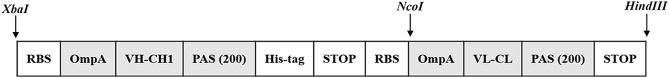
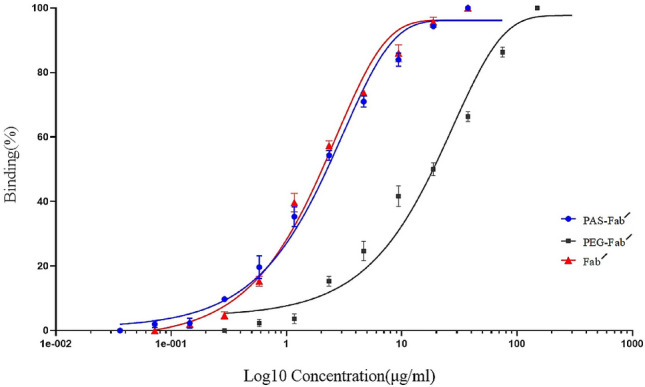
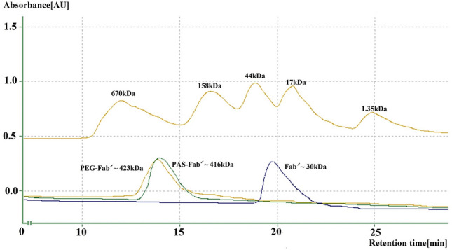
Certolizumab pegol is a Fab' antibody fragment for treatment of rheumatoid arthritis and Crohn's disease which is conjugated to a 40 kDa PEG molecule in order to increase the protein half-life. PEGylation may have disadvantages including immunogenicity, hypersensitivity, vacuolation, decreased binding affinity and biological activity of the protein. To overcome these problems, PASylation has been developed as a new approach. The nucleotide sequence encoding 400 amino acid PAS residues was genetically fused to the corresponding nucleotide sequences of both chains of certolizumab. Then, the bioactivity as well as physicochemical and pharmacokinetic properties of the recombinant PASylated expressed protein was assayed. Circular dichroism spectroscopy demonstrated that the random coil structure of PAS sequences did not change the secondary structure of the PASylated Fab' molecule. It was observed that PASylation influenced the properties of the Fab' molecule by which the hydrodynamic radius and neutralization activity were increased. Also, the antigen binding and binding kinetic parameters improved in comparison to the PEGylated Fab' antibody. Pharmacokinetic studies also showed prolonged terminal half-life and improved pharmacokinetic parameters in PASylated recombinant protein in comparison to the PEGylated and Fab' control molecules. The results reconfirmed the efficiency of PASylation approach as a potential alternative method in increasing the half-life of pharmaceutical proteins.


Conflict of interest statement

The authors declare no competing interests.

Figures

Figure 1
Figure 1
Final gene cassette expressing the PASylated Fab′ fragment in pET28a.
Figure 2
Figure 2
Analysis of protein purification process (non-reducing SDS-PAGE): 1: Eluted protein from one step Ni–NTA chromatography (~ 200 kDa band of PASylated Fab′ and ~ 110 kDa band of PASylated heavy chain); 2: Eluted protein from one step KappaSelect chromatography (~ 200 kDa band of PASylated Fab′ and ~ 110 kDa band of PASylated light chain); 3: Initial sample (IS) originated from the culture medium, M: Protein Mw marker, 4: Eluted ~ 200 kDa PASylated Fab′ from two-step purification procedure (KappaSelect followed by Ni–NTA affinity chromatography).
Figure 3
Figure 3
Analysis of protein purification process through Western blotting (non-reducing SDS-PAGE): 1: Eluted ~ 200 kDa PASylated Fab′ from two-step purification procedure (KappaSelect followed by Ni–NTA affinity chromatography); 2, 3: Eluted proteins from one step Ni–NTA chromatography (~ 200 kDa band of PASylated Fab′ and ~ 110 kDa band of PASylated heavy chain); M: Protein Mw marker. The image is cropped to show the one and two-step purification procedures.
Figure 4
Figure 4
Secondary structure analysis by Far-UV CD spectroscopy: Molar ellipticity calculations demonstrate a negative shift around 200 nm in the curve of PAS-Fab′ compared to its Fab′ antibody.
Figure 5
Figure 5
TNF-α ELISA binding activity of PAS-Fab′, PEG-Fab′ and Fab′ antibodies: data are represented as mean ± SD (three replicates).
Figure 6
Figure 6
RP-HPLC of PAS-Fab′ molecule in comparison to the corresponding Fab′ antibody: Samples were analyzed with gradient elution (2–80% CAN in 0.065–0.05% TFA) at 280 nm.
Figure 7
Figure 7
Size exclusion chromatography of PAS-Fab′, PEG-Fab′ and Fab′ antibodies: Mw marker includes the following proteins: Bovine thyroglobulin (670 kDa), bovine c-globulin (158 kDa), chicken ovalbumin (44 kDa), horse myoglobin (17 kDa), vitamin B12 (1.35 kDa).
Figure 8
Figure 8
Mass spectrometric analysis by MALDI-TOF/TOF: (a) A single peak with a molecular weight of 82,312.2 Da for non-reduced PAS-Fab′ antibody; (b) A single peak with a molecular weight of 48,013.7 Da for non-reduced Fab′ antibody.
Figure 9
Figure 9
Neutralization of TNF-α-mediated cytotoxicity in L929 cell line.
Figure 10
Figure 10
Thermo-analysis of tested PAS-Fab′, PEG-Fab′, and Fab′ antibodies: (a) DSC thermograms; (b) TG curves: samples were prepared in phosphate buffer (pH7.4) and analysis was performed at a scanning rate of 2 °C/min under nitrogen (inert) atmosphere. The two thermograms differed only to a negligible amount for PAS-Fab′ and Fab′ antibodies while PEG-Fab′ demonstrated a significant shift in comparison to the PAS-Fab′ and Fab′ antibodies regarded to the PEG molecule as a fusion. The melting curves of Fab′, PEG-Fab′, and PAS-Fab′ are highlighted in orange, blue, and green, respectively.
Figure 11
Figure 11
Murine pharmacokinetic profiles of PAS-Fab′, PEG-Fab′, and Fab′ antibodies.

References

    1. Holbrook J, Lara-Reyna S, Jarosz-Griffiths H, McDermott M. Tumour necrosis factor signaling in health and disease. F1000Res. 2019;8:271111. doi: 10.12688/f1000research.17023.1. - DOI - PMC - PubMed
    1. Mpofu S, Fatima F, Moots RJ. Anti-TNF therapies: They are all the same (aren’t they?) Rheumatology. 2005;44:271–273. doi: 10.1093/rheumatology/keh483. - DOI - PubMed
    1. Guo Q, et al. Rheumatoid arthritis: Pathological mechanisms and modern pharmacologic therapies. Bone Res. 2018;6:15. doi: 10.1038/s41413-018-0016-9. - DOI - PMC - PubMed
    1. Calabrò A, et al. One year in review 2016: Novelties in the treatment of rheumatoid arthritis. Clin. Exp. Rheumatol. 2016;34:357–372. - PubMed
    1. Shi HY, Ng SC. The state of the art on treatment of Crohn’s disease. J. Gastroenterol. 2018;53:989–998. doi: 10.1007/s00535-018-1479-6. - DOI - PMC - PubMed

Publication types