. 2020 Oct 28;10(1):18423.
doi: 10.1038/s41598-020-75451-5.

Deep phenotype unsupervised machine learning revealed the significance of pachychoroid features in etiology and visual prognosis of age-related macular degeneration

Affiliations

Deep phenotype unsupervised machine learning revealed the significance of pachychoroid features in etiology and visual prognosis of age-related macular degeneration

Yoshikatsu Hosoda et al. Sci Rep. .

Abstract

Unsupervised machine learning has received increased attention in clinical research because it allows researchers to identify novel and objective viewpoints for diseases with complex clinical characteristics. In this study, we applied a deep phenotyping method to classify Japanese patients with age-related macular degeneration (AMD), the leading cause of blindness in developed countries, showing high phenotypic heterogeneity. By applying unsupervised deep phenotype clustering, patients with AMD were classified into two groups. One of the groups had typical AMD features, whereas the other one showed the pachychoroid-related features that were recently identified as a potentially important factor in AMD pathogenesis. Based on these results, a scoring system for classification was established; a higher score was significantly associated with a rapid improvement in visual acuity after specific treatment. This needs to be validated in other datasets in the future. In conclusion, the current study demonstrates the usefulness of unsupervised classification and provides important knowledge for future AMD studies.


Conflict of interest statement

The authors declare no competing interests.

Figures

Figure 1
Figure 1
Examples of clinical characteristics. (AC) Images from CNV fellow eye of a 79 year old male patient. (A) Color fundus photograph. Type A drusen in the macular region (blue arrow). (B) Late phase of ICGA image. (C) Foveal horizontal OCT scan shows choroidal thinning. (DF) Images from CNV fellow eye of a 64 year old male patient. (D) Color fundus photograph shows multiple type B and C drusen within and outside macular region (blue arrows). (E) Choroidal hyperpermeability in late phase of ICGA (blue arrows). (F) Foveal horizontal EDI OCT scan shows choroidal thickening and dilated choroidal vessels (blue arrows). (GI) Images from CNV fellow eye of an 87 year old female patient. (G) Color fundus photograph shows type A drusen (red arrow) and pseudodrusen (blue arrow) within macular region. (H) Late phase of ICGA. (I) Foveal horizontal OCT scan shows the accumulation of deposits on retinal pigment epithelium (blue arrows). CNV choroidal neovascularization, EDI enhanced depth imaging, ICGA indocyanine green angiography, OCT optical coherence tomography.
Figure 2
Figure 2
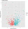
Scatter plot showing the result of machine learning clustering. Unsupervised machine learning algorithm was used to cluster patient data. The scatter plot indicates the position of the subjects according to the similarity by dimensionality reduction. Patients were divided into cluster 1 (red dots, N = 289) and cluster 2 (blue dots, N = 247).
Figure 3
Figure 3
Histograms of subfoveal choroidal thickness in CNV-affected eye (A) and fellow eye (B). Superposed histograms of patients in PNV-type cluster (blue) and AMD-type cluster (red) show binominal distribution of subfoveal choroidal thickness (SFCT) in both eyes. CNV; choroidal neovascularization.
Figure 4
Figure 4
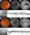
A case of low CNV score corresponding to AMD-type patient. A 76-year-old male patient was visually impaired in the right eye. Images from the right eye (AD) and left eye (EI). (A) Color fundus photograph shows large retinal pigment epithelium detachment. (B) Fluorescein angiography showed occult CNV suggesting type 1 CNV. (C) Late phase of indocyanine green angiography image shows no choroidal vascular hyperpermeability. (D) Infrared reflectance image of right eye. (E) Foveal horizontal OCT scan shows large pigment epithelial detachment consistent with CNV lesion. Subfoveal choroidal thickness was 128 μm and retinal thickness was 1197 μm. (F) Color fundus photograph shows type A drusen within macular lesion (blue arrow). There are no type B drusen or type C drusen. (G) Fluorescein angiography image suggests no CNV. (H) Late phase of indocyanine green angiography image shows no choroidal vascular hyperpermeability. (I) Infrared reflectance image of left eye. (J) Foveal horizontal OCT scan shows a subretinal deposit consistent with type A drusen. Subfoveal choroidal thickness was 145 μm and retinal thickness was 252 μm. CNV score of this patient was − 7.978, indicating AMD. CNV choroidal neovascularization, OCT optical coherence tomography, AMD age-related macular degeneration.
Figure 5
Figure 5
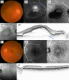
A case of high CNV score corresponding to PNV-type patient. A 66-year-old male patient was visually impaired in the right eye. Images from the right eye (AD) and left eye (EI). (A) Color fundus photograph shows subretinal hemorrhage within macular region (blue arrow). (B) Fluorescein angiography image shows leakage within the region of subretinal hemorrhage. (C) Late phase of indocyanine green angiography image shows a polypoidal lesion (blue arrow). Choroidal vascular hyperpermeability spots were also observed around the macular region. (D) Infrared reflectance image of right eye. (E) Foveal vertical EDI OCT scan shows diffusely thickened choroid and pigment epithelial detachment. Dilated choroidal vessels were also observed (blue arrows). Subfoveal choroidal thickness was 508 μm and retinal thickness was 455 μm. (F) Color fundus photograph shows no CNV. (G) Fluorescein angiography image shows no leakage. (H) Late phase of indocyanine green angiography image shows multiple choroidal vascular hyperpermeability spots (blue arrows). (I) Infrared reflectance image of left eye. (J) Foveal horizontal EDI OCT scan shows diffusely thickened choroid and dilated choroidal vessels (blue arrows). Subfoveal choroidal thickness was 503 μm and retinal thickness was 262 μm. CNV score for this patient was 7.656, which indicating PNV. CNV choroidal neovascularization, EDI enhanced depth imaging, OCT optical coherence tomography, PNV pachychoroid neovasculopathy.

References

    1. Smith W, et al. Risk factors for age-related macular degeneration: pooled findings from three continents. Ophthalmology. 2001;108:697–704. doi: 10.1016/S0161-6420(00)00580-7. - DOI - PubMed
    1. Bressler NM, et al. Potential public health impact of age-related eye disease study results: AREDS Report No. 11. Arch. Ophthalmol. 2003;121:1621–1624. doi: 10.1001/archopht.121.1.113. - DOI - PMC - PubMed
    1. Hageman GS, et al. An integrated hypothesis that considers drusen as biomarkers of immune-mediated processes at the RPE-Bruch’s membrane interface in aging and age-related macular degeneration. Prog. Retin. Eye Res. 2001;20:705–732. doi: 10.1016/S1350-9462(01)00010-6. - DOI - PubMed
    1. Johnson LV, Leitner WP, Staples MK, Anderson DH. Complement activation and inflammatory processes in drusen formation and age related macular degeneration. Exp. Eye Res. 2001;73:887–896. doi: 10.1006/exer.2001.1094. - DOI - PubMed
    1. Scholl S, Kirchhof J, Augustin AJ. Role of inflammation in the pathogenesis of age-related macular degeneration. Expert Rev. Ophthalmol. 2009;4:617–625. doi: 10.1586/eop.09.51. - DOI

MeSH terms

Substances