. 2020 Jun:44:67-97.
doi: 10.3767/persoonia.2020.44.03. Epub 2019 Aug 29.

A new species concept for the clinically relevant Mucor circinelloides complex

Affiliations

A new species concept for the clinically relevant Mucor circinelloides complex

L Wagner et al. Persoonia. 2020 Jun.

Abstract

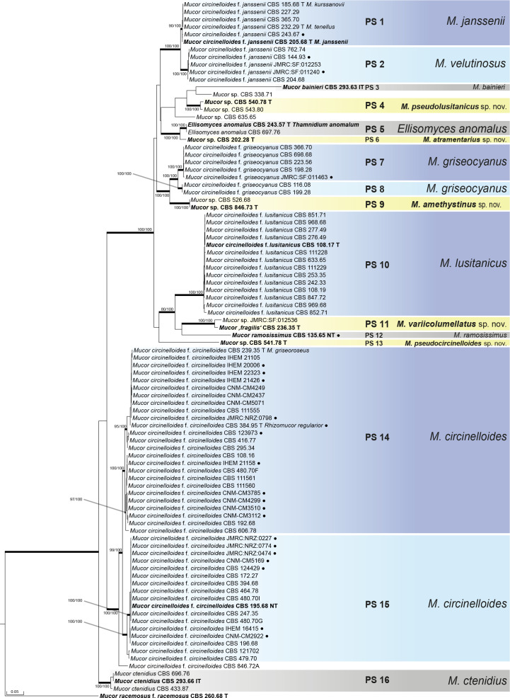
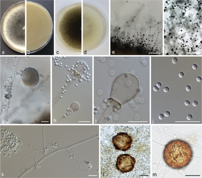
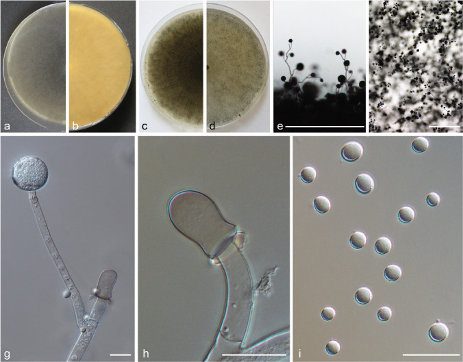
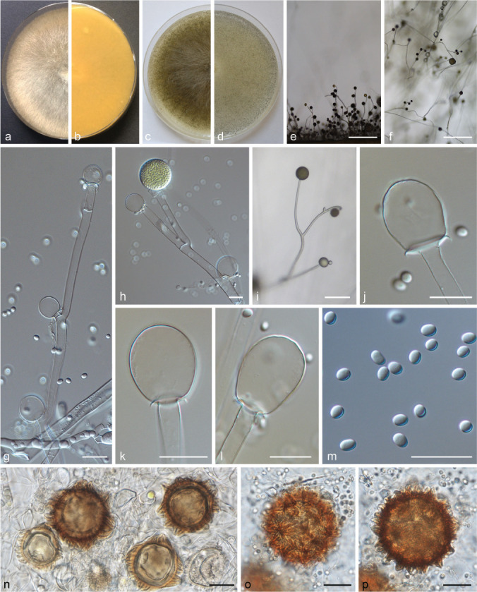
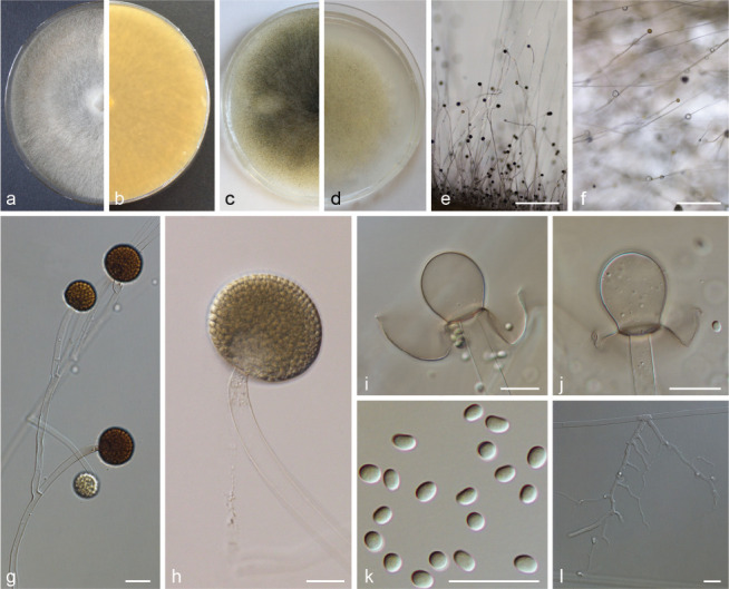
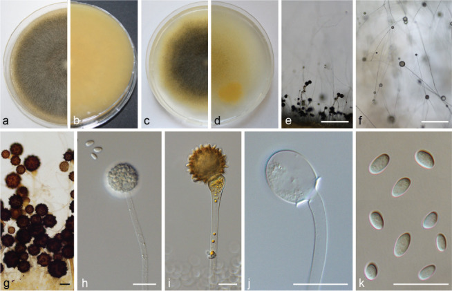
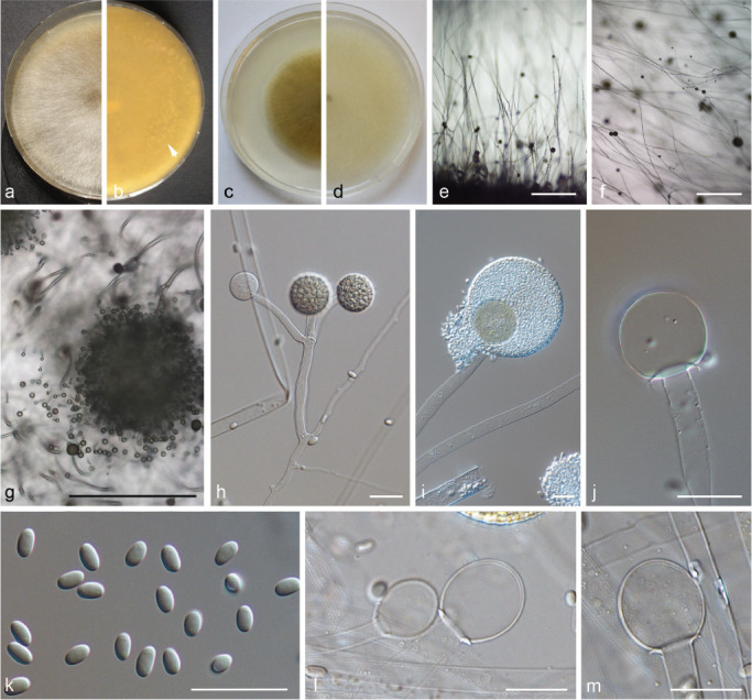
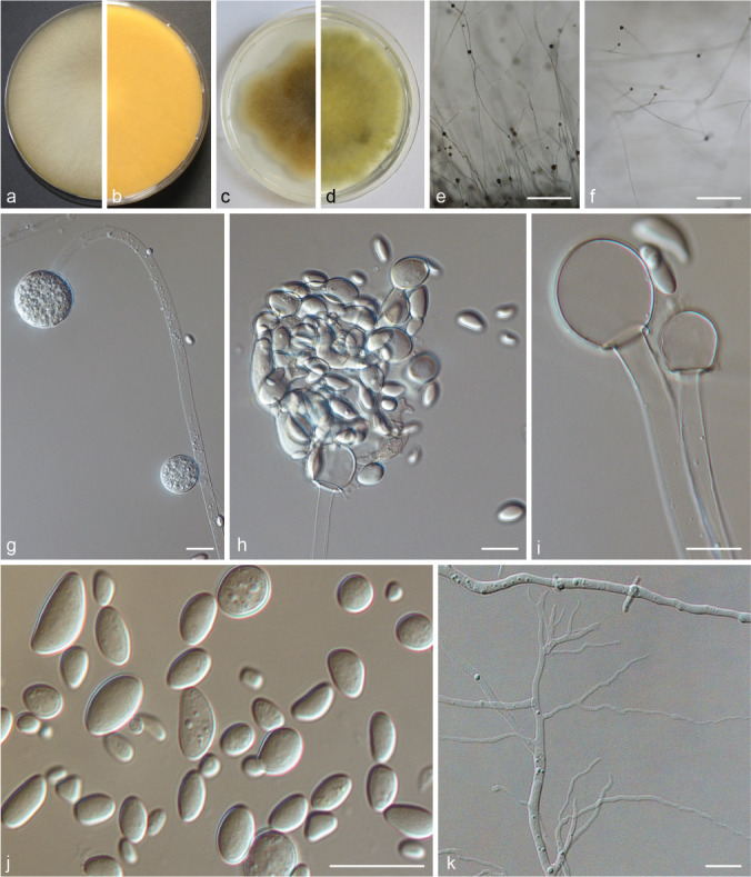
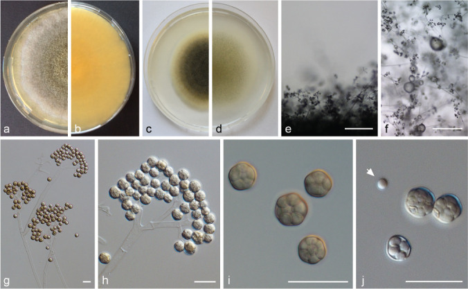
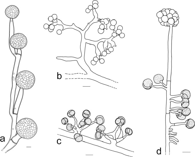
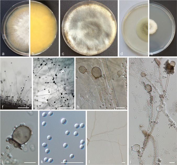
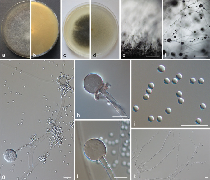
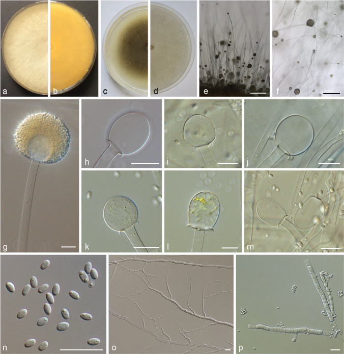
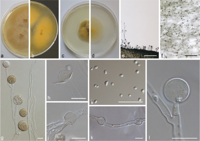
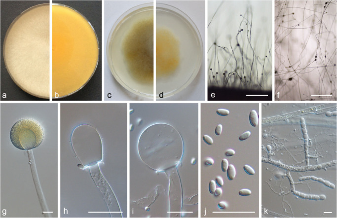
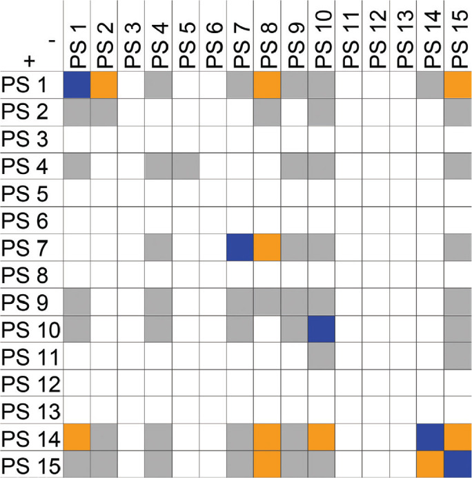
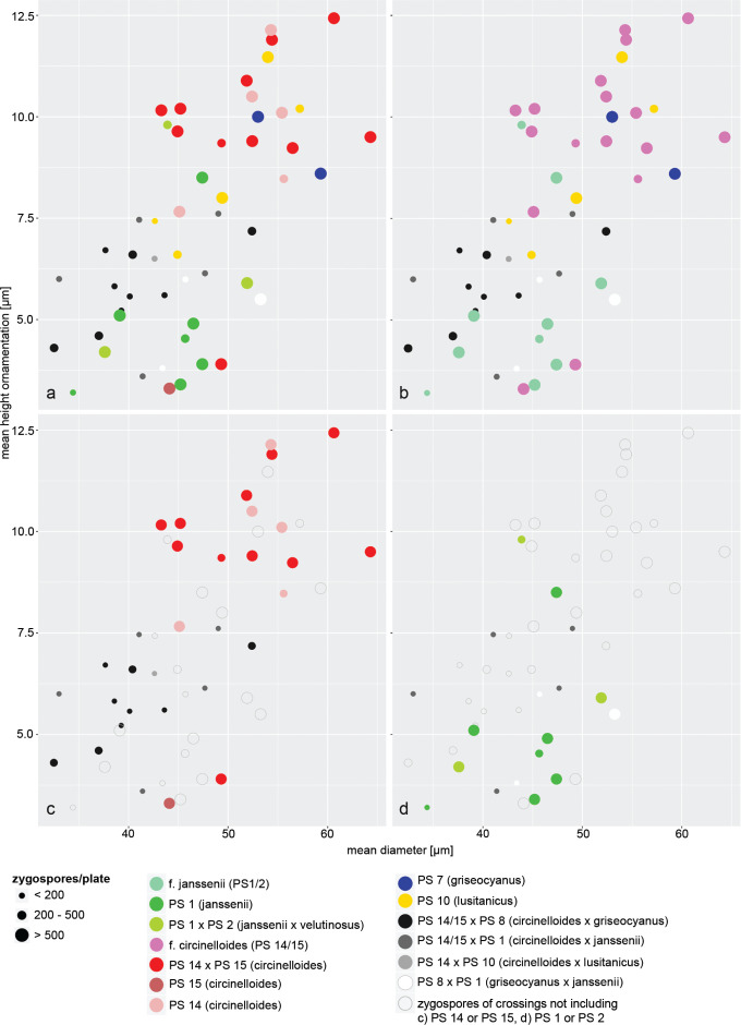
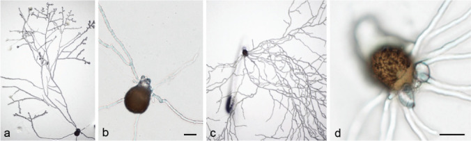
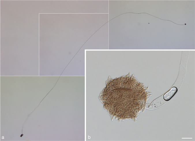
Mucor species are common soil fungi but also known as agents of human infections (mucormycosis) and used in food production and biotechnology. Mucor circinelloides is the Mucor species that is most frequently isolated from clinical sources. The taxonomy of Mucor circinelloides and its close relatives (Mucor circinelloides complex - MCC) is still based on morphology and mating behaviour. The aim of the present study was a revised taxonomy of the MCC using a polyphasic approach. Using a set of 100 strains molecular phylogenetic analysis of five markers (ITS, rpb1, tsr1, mcm7, and cfs, introduced here) were performed, combined with phenotypic studies, mating tests and the determination of the maximum growth temperatures. The multi-locus analyses revealed 16 phylogenetic species of which 14 showed distinct phenotypical traits and were recognised as discrete species. Five of these species are introduced as novel taxa: M. amethystinus sp. nov., M. atramentarius sp. nov., M. variicolumellatus sp. nov., M. pseudocircinelloides sp. nov., and M. pseudolusitanicus sp. nov. The former formae of M. circinelloides represent one or two separate species. In the MCC, the simple presence of well-shaped zygospores only indicates a close relation of both strains, but not necessarily conspecificity. Seven species of the MCC have been implemented in human infection: M. circinelloides, M. griseocyanus, M. janssenii, M. lusitanicus, M. ramosissimus, M. variicolumellatus, and M. velutinosus.

Keywords: Mucor; mating tests; maximum growth temperature; mucormycosis; new taxa; phylogeny; taxonomy; zygospore formation.


Figures

Fig. 1
Fig. 1
Gene model of the putative cyclopropane-fatty-acyl-phospholipid synthase following the gene prediction (JGI, estExt_Genewise1Plus.C_011066 of Mucor circinelloides CBS277.49 genome v2.0) with primer binding sites and estimated amplicon length.
Fig. 2
Fig. 2
Phylogenetic tree based on the concatenated alignment of five molecular markers (cfs, mcm7, ITS, rpb1, tsr1) calculated using RAxML. Branches with bootstrap values ≥ 75 in the RAxML and/or the Maximum Parsimony analyses are printed in bold and the bootstrap values are given, in this order. T = ex-type strain, IT = ex-isotype strain and NT = ex-neotype strain. Ex-type strains of currently accepted taxa are printed in bold. Clinical isolates are marked by black circles.
Fig. 3
Fig. 3
Columellae of the M. racemosus group: a–g. MCC, typical columellae of tall sporangiophores without or only with slightly truncate bases (a–b. M. circinelloides CBS 195.68; c. M. circinelloides CBS 384.95; d–g. M. janssenii CBS 205.68); h–o. MCC columellae of short sporangiophores of M. janssenii and M. velutinosus (h–l. M. velutinosus JMRC:SF:011240; m–o. M. janssenii CBS 185.68); p–w. MRC, typical columellae (p–q. M. racemosus f. racemosus CBS 260.68; r–t. M. racemosus f. racemosus CBS 774.86; u–v. M. racemosus f. sphaerosporus CBS 115.08; w. M. plumbeus CBS 226.36).
Fig. 4
Fig. 4
PS 1 (M. janssenii) CBS 205.68. a. Front and b. reverse of a 7-d-old culture on MEA at room temperature (RT); c. front on PDA; d. front on SMA; e. lateral view; f. top view; g. sporangium; h–i. columellae with collar; j. sporangiospores; k. rhizoids; l–m. zygospores of crosses with M. griseocyanus CBS 116.08. — Scale bars: e–f = 500 μm; g–m = 20 μm.
Fig. 5
Fig. 5
PS 2 (M. velutinosus) CBS 762.74. a. Front and b. reverse of a 7-d-old culture on MEA at RT; c. front on PDA; d. front on SMA; e. lateral view; f. top view; g. sporangiophore with sporangium and columella; h. columella with collar; i. sporangiospores. — Scale bars: e–f = 500 μm; g–i = 20 μm.
Fig. 6
Fig. 6
PS 14/15 (M. circinelloides) (a–h, k–m. CBS 195.68; i–j. CBS 384.95). a. Front and b. reverse of a 7-d-old culture on MEA at RT; c. front on PDA; d. front on SMA; e. lateral view; f. top view; g. sporangiophore with columellae; h. sporangiophore with sporangium and columella; i. sporangiophore with sporangia; j–l. columella; m. sporangiospores; n. zygospore formed in crosses of CBS 192.68 × CBS 124429; o–p. zygospores formed in crosses of CBS 192.68 × CBS 394.68. — Scale bars: e–f = 500 μm; g–p = 20 μm.
Fig. 7
Fig. 7
PS 7 (M. griseocyanus) CBS 366.70. a. Front and b. reverse of a 7-d-old culture on MEA at R; c. front on PDA; d. front on SMA; e. lateral view; f. top view; g. sporangiophore with sporangia; h. sporangium; i–j. columella with collar; k. sporangiospores; l. rhizoids. — Scale bars: e–f = 500 μm; g–l = 20 μm.
Fig. 8
Fig. 8
PS 3 (M. bainieri) CBS 293.63. a. Front and b. reverse of a 7-d-old culture on MEA at RT; c. front on PDA; d. front on SMA; e. lateral view; f. top view; g. lateral view with azygospores; h. sporangium; i. azygospore; j. columella with collar; k. sporangiospores. — Scale bars: e–f = 500 μm; g–k = 20 μm.
Fig. 9
Fig. 9
PS 4 (M. pseudolusitanicus sp. nov.) CBS 540.78. a. Front and b. reverse with protuberances (arrow) of a 7-d-old culture on MEA at RT; c. front on PDA; d. front on SMA; e. lateral view; f. top view; g. top view on protuberance; h. sporangiophore with sporangia; i. sporangium; j. columella; k. sporangiospores; l–m. columellae of CBS 635.35. — Scale bars: e–f = 500 μm; g–m = 20 μm.
Fig. 10
Fig. 10
PS 10 (M. lusitanicus) CBS 108.17. a. Front and b. reverse of a 7-d-old culture on MEA at RT; c. front on PDA; d. front on SMA; e. lateral view; f. top view; g. sporangiophore with sporangia; h. burst sporangium releasing sporangiospores; i. columellae; j. sporangiospores; k. rhizoids. — Scale bars: e–f = 500 μm; g–k = 20 μm.
Fig. 11
Fig. 11
PS 5 (Ellisomyces anomalus) CBS 243.57. a. Front and b. reverse of a 7-d-old culture on MEA at RT; c. front on PDA; d. front on SMA; e. lateral view; f. top view; g. sporophore with sporangiola; h. sporangiola on sporophore; i. sporangiola; j. sporangiola and single spore (arrow) — Scale bars: e–f = 500 μm; g–j = 20 μm.
Fig. 12
Fig. 12
Sporophores in the MCC. a. M. ramosissimus CBS 135.65; b. Ellisomyces anomalus CBS 243.37 (young sporophore); c. M. ctenidius CBS 433.87; d. M. ctenidius CBS 293.66. — Scale bars = 10 μm.
Fig. 13
Fig. 13
PS 6 (M. atramentarius sp. nov.) CBS 202.28. a. Front and b. reverse of a 7-d-old culture on MEA at RT; c. front view of a 3 weeks old culture on MEA; d. 7-d-old culture on PDA and e. front on SMA; f. lateral view; g. top view; h. sporangiophore and columellae; i. sporangiophore; j. columella; k. sporangiospores; l. rhizoids. — Scale bars: f–g = 500 μm; h–l = 20 μm.
Fig. 14
Fig. 14
PS 9 (M. amethystinus sp. nov.) CBS 846.73. a. Front and b. reverse of a 7-d-old culture on MEA at RT; c. front on PDA; d. front on SMA; e. lateral view; f. top view; g. side branch of a sporangiophore; h–i. columella with collar; j. sporangiospores; k. rhizoids. — Scale bars: e–f = 500 μm; g–k = 20 μm.
Fig. 15
Fig. 15
PS 11 (M. variicolumellatus sp. nov.) CBS 236.35 and JMRC:SF:012536. a. Front and b. reverse of a 7-d-old culture on MEA at RT; c. front on PDA; d. front on SMA; e. lateral view; f. top view; g. sporangium with translucent columella; h–m. columellae; n. sporangiospores; o. rhizoid; p. chlamydospores. — Scale bars: e–f = 500 μm; g–p = 20 μm.
Fig. 16
Fig. 16
PS 12 (M. ramosissiumus) CBS 135.65. a. Front and b. reverse of a 7-d-old culture on MEA at RT; c. front on PDA; d. front on SMA; e. lateral view; f. top view; g. sporangiophore with sporangia; h, j. columellae with collar; i. sporangiospores; k. hyphal bulges; l. sterile sporangium. — Scale bars: e–f = 500 μm; g–l = 20 μm.
Fig. 17
Fig. 17
PS 13 (M. pseudocircinelloides sp. nov.) CBS 541.78. a. Front and b. reverse of a 7-d-old culture on MEA at RT; c. front on PDA; d. front on SMA; e. lateral view; f. top view; g. sporangium with translucent columella; h–i. columella; j. sporangiospores; k. rhizoids and chlamydospores. — Scale bars: e–f = 500 μm; g–k = 20 μm.
Fig. 18
Fig. 18
PS 16 (M. ctenidius) CBS 293.66. a. Front and b. reverse of a 7-d-old culture on MEA at RT; c. front on PDA; d. front on SMA; e. lateral view; f. top view; g–h. sporangiophore with terminal sporangium; i. columella; j–k. sporangiola; l. sporangiola of CBS 433.87 with columella; m. sporangiola. — Scale bars: e–f = 500 μm; g–m = 20 μm.
Fig. 19
Fig. 19
Zygospores of the MCC. a–d. Well-developed zygospores (a–c. zygospores of intraformae cross within f. circinelloides CBS 394.68 × CBS 172.27: c. different states of zygospore development (gametangium, young zygospore, maturing zygospore, matured zygospore); d. zygospores of intraformae cross within f. circinelloides CBS 394.68 × CBS 192.68); e–f. not well-developed zygospores (e. interformae cross between f. griseocyanus CBS 116.08 and f. circinelloides CNM-CM3112; f. zygospores of interformae cross between f. griseocyanus CBS 116.08 and f. janssenii CBS 205.68); g. gametangia of interformae cross between f. griseocyanus CBS 116.08 and f. janssenii CBS 205.68. — Scale bars = 50 μm.
Fig. 20
Fig. 20
Overview of performed crosses. PS arranged according to their mating types (+/–): orange – formation of inter PS zygospores, blue – formation of intra PS zygospores, grey – cross without formation of zygospores, white – no cross performed.
Fig. 21
Fig. 21
Size, height of ornamentation and abundance of zygospores formed in the MCC. a. Intra- and inter PS crosses; b. intra- and interformae crosses; c. intra- and inter PS crosses (PS 14 × PS 15) of f. circinelloides; d. intra- and inter PS crosses (PS 1, M. janssenii × PS 2, M. velutinosus) of f. janssenii.
Fig. 22
Fig. 22
a–d. Mycelial germination of zygospores of crosses within f. circinelloides (PS 15, CBS 394.68 × CBS 172.27). — Scale bars = 20 μm.
Fig. 23
Fig. 23
Sporangial germination of a zygospore of crosses within f. circinelloides (PS 15, CBS 394.68 × CBS 172.27). a. Germ sporangiophore, total height about 1 cm (photographs assembled); b. zygospore with basal part of germ sporangiophore. — Scale bar = 20 μm.

References

    1. Abe A, Asano K, Sone T. 2010. A molecular phylogeny-based taxonomy of the genus Rhizopus. Bioscience, Biotechnology, and Biochemistry 74: 1325–1331. - PubMed
    1. Aguileta G, Marthey S, Chiapello H, et al. 2008. Assessing the performance of single-copy genes for recovering robust phylogenies. Systematic Biology 57: 613–627. - PubMed
    1. Alastruey-Izquierdo A, Hoffmann K, De Hoog GS, et al. 2010. Species recognition and clinical relevance of the zygomycetous genus Lichtheimia (syn. Absidia pro parte, Mycocladus). Journal of Clinical Microbiology 48: 2154–2170. - PMC - PubMed
    1. Álvarez E, Cano J, Stchigel AM, et al. 2011. Two new species of Mucor from clinical samples. Medical Mycology 49: 62–72. - PubMed
    1. Álvarez E, Stchigel AM, Cano J, et al. 2010. Molecular phylogenetic diversity of the emerging mucoralean fungus Apophysomyces: proposal of three new species. Revista Iberoamericana de Micología 27: 80–89. - PubMed

LinkOut - more resources