. 2020 Sep 29:11:1997.
doi: 10.3389/fmicb.2020.01997. eCollection 2020.

Mass Spectrometric Characterization of HSV-1 L-Particles From Human Dendritic Cells and BHK21 Cells and Analysis of Their Functional Role

Affiliations

Mass Spectrometric Characterization of HSV-1 L-Particles From Human Dendritic Cells and BHK21 Cells and Analysis of Their Functional Role

Alexandra Birzer et al. Front Microbiol. .

Abstract

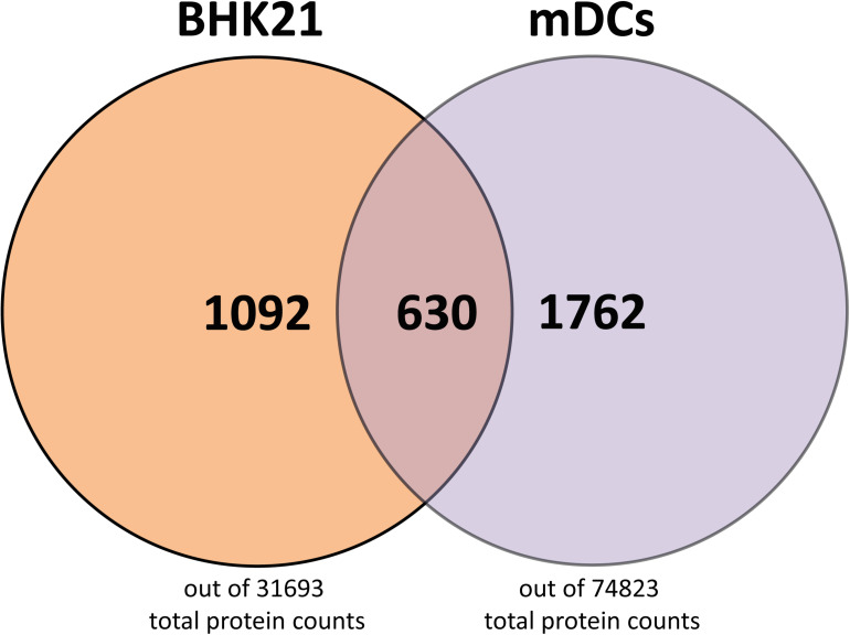
Herpes simplex virus type 1 (HSV-1) is a very common human pathogenic virus among the world's population. The lytic replication cycle of HSV-1 is, amongst others, characterized by a tripartite viral gene expression cascade, the assembly of nucleocapsids involving their subsequent nuclear egress, tegumentation, re-envelopment and the final release of progeny viral particles. During productive infection of a multitude of different cell types, HSV-1 generates not only infectious heavy (H-) particles, but also non-infectious light (L-) particles, lacking the capsid. In monocyte-derived mature dendritic cells (mDCs), HSV-1 causes a non-productive infection with the predominant release of L-particles. Until now, the generation and function of L-particles is not well understood, however, they are described as factors transferring viral components to the cellular microenvironment. To obtain deeper insights into the L-particle composition, we performed a mass-spectrometry-based analysis of L-particles derived from HSV-1-infected mDCs or BHK21 cells and H-particles from the latter one. In total, we detected 63 viral proteins in both H- and L-particle preparations derived from HSV-1-infected BHK21 cells. In L-particles from HSV-1-infected mDCs we identified 41 viral proteins which are differentially distributed compared to L-particles from BHK21 cells. In this study, we present data suggesting that L-particles modify mDCs and suppress their T cell stimulatory capacity. Due to the plethora of specific viral proteins incorporated into and transmitted by L-particles, it is tempting to speculate that L-particles manipulate non-infected bystander cells for the benefit of the virus.

Keywords: BHK21 cells; HSV-1; T cell stimulation; dendritic cells; heavy particles; immunomodulatory effect; light particles; mass spectrometry.


Figures

FIGURE 1
FIGURE 1
Purification of L- and H-particles derived from HSV-1-infected BHK21 cells or mDCs. L-particles and H-particles were purified from supernatants of HSV-1-infected BHK21 cells and mDCs four days post infection and 20 hpi, respectively. (A) Representative Ficoll gradients from HSV-1-infected BHK21 cells (left panel) and mDCs (right panel) are shown. (B) Western blot analyses of L- and H-particle protein lysates derived from BHK21 cells. A total protein amount of approximately 4 μg was loaded onto a 10% SDS gel. (C) Western blot analyses of L-particle protein lysates derived from HSV-1-infected mDCs (left panel) in comparison to BHK21 cell-derived H-particles (right panel). (B,C) Purity of particle preparations was verified by detection of ICP5, ICP4, ICP0, and gB using specific antibodies. Exposure time for detection is depicted as seconds (s). (D) Coomassie blue staining of H- and L-particle samples isolated from HSV-1-infected BHK21 cells loaded onto a 10% SDS-PAA gel (left). Silver staining of L-particles derived from HSV-1-infected mDCs loaded onto a 10% SDS-PAA gel (right). (E) Electron microscopic analyses of HSV-1 particles derived from either BHK21 cells or mDCs. HSV-1 particles were adhered to carbon-coated grids and stained using 1% uranyl acetate and lead citrate. Bars, 250 nm (particles derived from BHK21 cells) and 500 nm (L-particles derived from mDCs).
FIGURE 2
FIGURE 2
Viral protein distribution of HSV-1-derived particles isolated from infected BHK21 cells or mDCs. (A,B) L-particles and H-particles were purified from supernatants of HSV–1-infected BHK21 cells (A) and mDCs (B) four days post infection and 20 hpi, respectively. Protein distribution of L- and H-particles was analyzed by mass spectrometry. The total intensity of viral proteins was set to 100% and relative values (normalized to the mean of all present glycoproteins) for H- and L-particle samples are shown regarding their predicted protein localization. The data are based on four BHK21cell- or three mDC-derived particle preparations.
FIGURE 3
FIGURE 3
Mass spectrometry-based comparative analysis of HSV-1-derived L-particles versus H-particles from infected BHK21 cells. HSV–1-derived H- and L-particles from supernatants of infected BHK21 cells were isolated for subsequent mass spectrometric analysis. The results of all four replicates are shown as relative ratio of L- versus H-particles using the respective protein intensity normalized to the mean intensity of all detected glycoproteins. Proteins are grouped according to their annotation as envelope (A), tegument (B), non-structural (C) or capsid proteins (D) and ordered regarding their ratio. The dashed red line represents a ratio of 1. UL3 was detected in two out of three samples and is marked with “#.” The values in the heat maps represent the average intensity of each detected protein among different samples, normalized to the mean of all detected glycoproteins, in the respective samples. Normalized values were separately calculated for L- and H-particle samples.
FIGURE 4
FIGURE 4
Mass spectrometry-based analysis of HSV-1-derived L-particles from mDCs. HSV–1-derived L-particles were isolated from supernatants of infected mDCs and used for subsequent mass spectrometric analysis. The results of all three replicates are shown as individual intensities normalized to the mean intensity of all glycoproteins in the respective sample (heat map, graph). Proteins are grouped according to their annotation as envelope (A), tegument (B), non-structural (C) or capsid proteins (D). Proteins detected in two out of three replicates are marked with “#.”
FIGURE 5
FIGURE 5
Schematic protein signature of HSV-1 L-particles derived from HSV-1-infected BHK21 cells and mDCs. Graphical comparison of the viral protein signature of HSV-1 L-particles derived from infected BHK21 cells (left part) and mDCs (right part) based on the mean intensity of all samples (BHK21: four samples, mDCs: three samples) as shown in Tables 1, 2. The top abundant hits are shown as circles, each representing a single viral protein. The diameter was adapted according to the relative signal intensities of the depicted viral proteins. The proteins are categorized regarding their predicted localization within the virion, i.e., tegument (black), envelope (gray), capsid (blue), or non-structural proteins (green).
FIGURE 6
FIGURE 6
Host cell proteins in L- and H-particles derived from BHK21 cell and L-particles derived from mDCs. A total number of host cell proteins of 1092 were found in H- and L-particles from HSV-1-infected BH21 cells, whereas 1762 host cell proteins were detected in L-particles derived from HSV-1-infected mDCs. 630 host cell proteins were identical in the HSV-1 particles derived from these different cell types. Data is evaluated based on the databases from Mesocricetus auratus (BHK21 cells) and Homo sapiens (mDCs).
FIGURE 7
FIGURE 7
L-particles selectively interfere with CD83 surface expression during DC maturation. Immature DCs (iDCs) were infected with HSV-1 (MOI of 2), treated with UV-inactivated HSV-1 (viral material corresponding to MOI of 20, 8 × 0.12 J/cm2) or L-particles (viral material corresponding to high MOI, 3 × 0.12 J/cm2) or left untreated. At 1hpi, infected cells were transferred into medium containing a defined maturation cocktail. An uninfected iDC sample served as an input control and was directly stained against CD11c, CD80, CD83, CCR7, and MHC class II (MHCII) for flow cytometric analyses. Cells were harvested 24 hpi, stained with specific antibodies mentioned before and used for flow cytometry. The experiment was performed five times with cells from different healthy donors. Error bars indicate SEM. Significant changes to mock were analyzed using a one-way ANOVA and Bonferroni multiple comparison post hoc tests and are indicated by asterisks (***indicates p ≤ 0.001 and ****p ≤ 0.0001) and values depicted as “ns” were not significant (p < 0.05).
FIGURE 8
FIGURE 8
L-particles possess an immunomodulatory effect on mDCs. Mature DCs (mDCs) were infected with purified H-particles (MOI of 2), treated with UV–irradiated H-particles (viral material corresponding to MOI of 20, 8 × 0.12 J/cm2) or L-particles (viral material corresponding to high MOI, 3 × 0.12 J/cm2) or left untreated (mock). At 8 hpi, cells were cocultured with allogeneic T cells in a mixed lymphocyte reaction (MLR) for additional 72 h. Cells were pulsed with 1 μCi/well [3H]-thymidine (PerkinElmer) for 16 h before harvesting. The experiment was performed three times with mDCs from different healthy donors. Error bars indicate SEM. Statistical analyses was performed relative to mock values and is depicted on the right side. Only the significant changes to mock were analyzed using a one-way ANOVA and Bonferroni multiple comparison post hoc tests and only significant changes are indicated by asterisks (*indicates p ≤ 0.05, **p ≤ 0.01, ***p ≤ 0.001, and ****p ≤ 0.0001).

References

    1. Alandijany T. (2019). Host intrinsic and innate intracellular immunity during herpes simplex Virus Type 1 (HSV-1) Infection. Front. Microbiol. 10:2611. 10.3389/fmicb.2019.02611 - DOI - PMC - PubMed
    1. Alemañ N., Quiroga M. I., López-Peña M., Vázquez S., Guerrero F. H., Nieto J. M. (2003). L-particle production during primary replication of pseudorabies virus in the nasal mucosa of swine. J. Virol. 77 5657–5667. 10.1128/jvi.77.10.5657-5667.2003 - DOI - PMC - PubMed
    1. Alvarez D., Vollmann E. H., von Andrian U. H. (2008). Mechanisms and consequences of dendritic cell migration. Immunity 29 325–342. 10.1016/j.immuni.2008.08.006 - DOI - PMC - PubMed
    1. Bantscheff M., Schirle M., Sweetman G., Rick J., Kuster B. (2007). Quantitative mass spectrometry in proteomics: a critical review. Anal. Bioanal. Chem. 389 1017–1031. 10.1007/s00216-007-1486-6 - DOI - PubMed
    1. Bigalke J. M., Heldwein E. E. (2017). Have NEC coat, will travel: structural basis of membrane budding during nuclear egress in herpesviruses. Adv. Virus Res. 97 107–141. 10.1016/bs.aivir.2016.07.002 - DOI - PMC - PubMed