. 2021 Jan;105(1):223-244.
doi: 10.1111/tpj.15053. Epub 2020 Nov 27.

Developmental acclimation of the thylakoid proteome to light intensity in Arabidopsis

Affiliations

Developmental acclimation of the thylakoid proteome to light intensity in Arabidopsis

Sarah E Flannery et al. Plant J. 2021 Jan.

Abstract

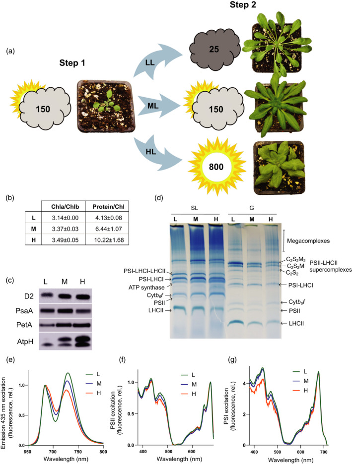
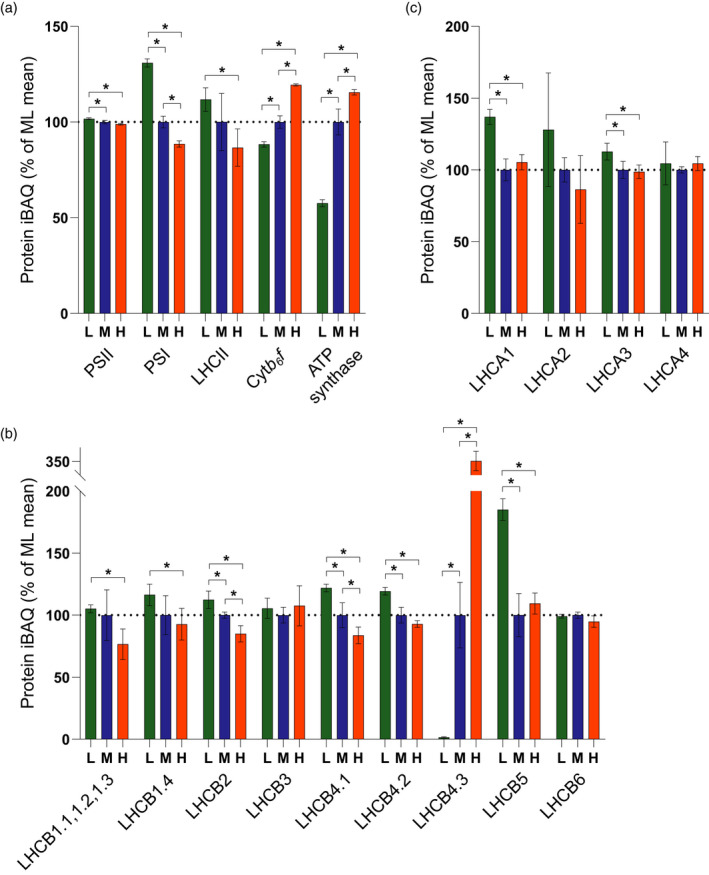
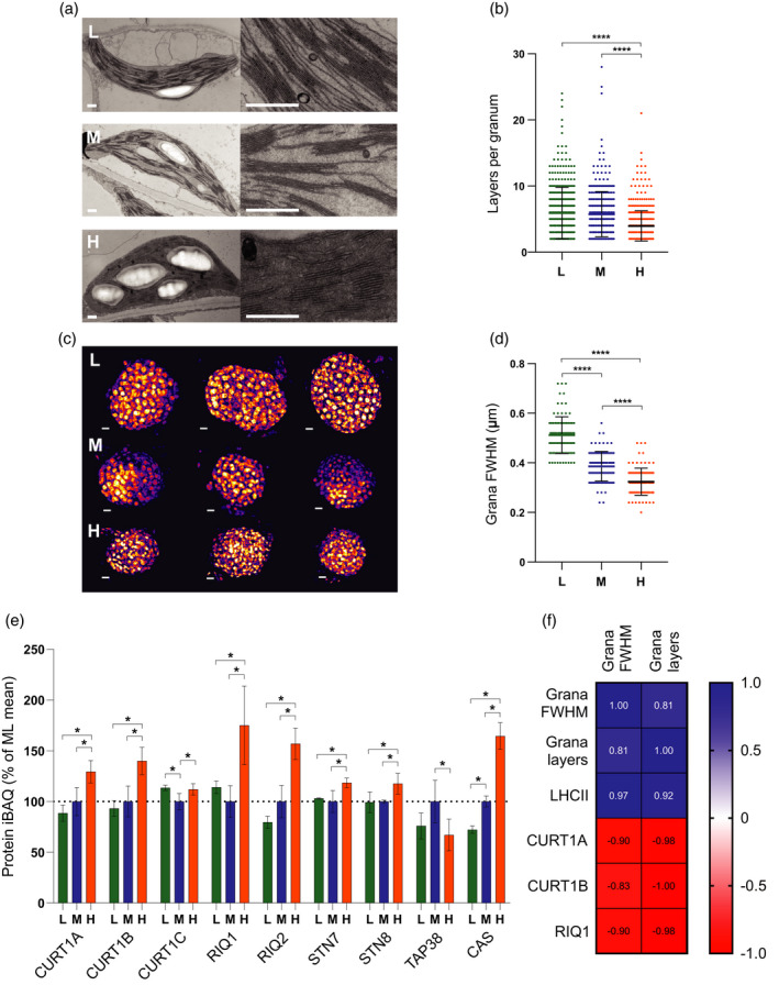
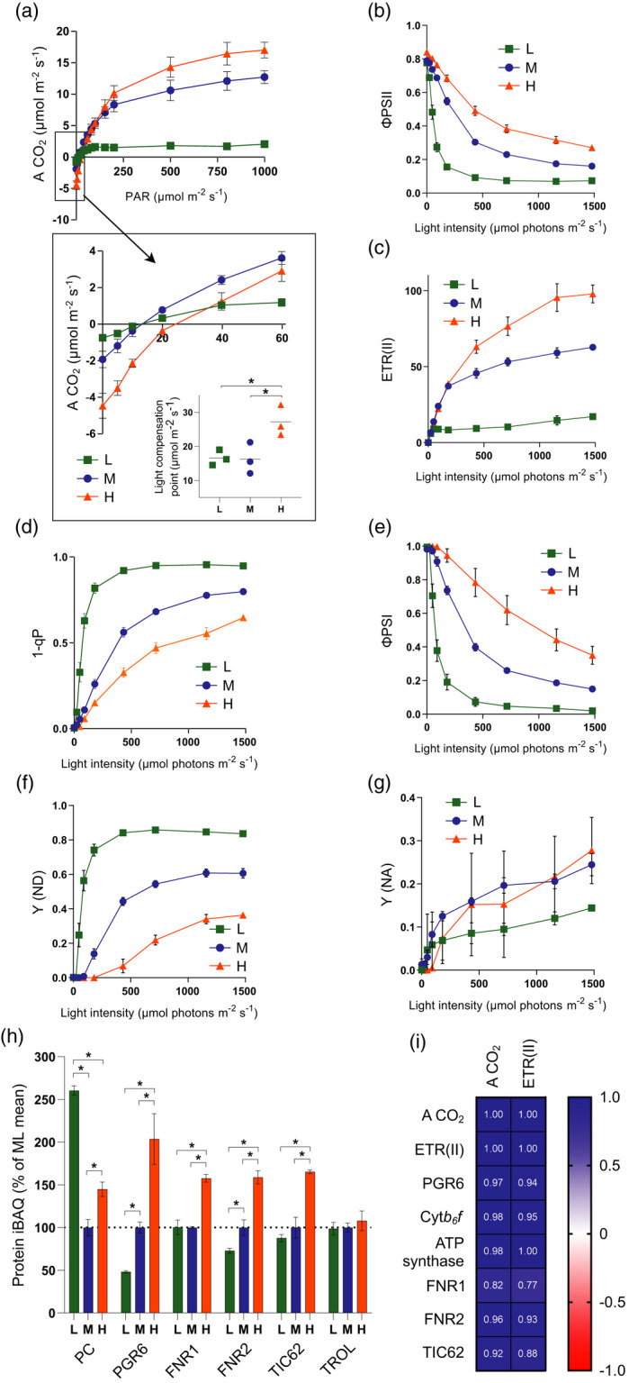
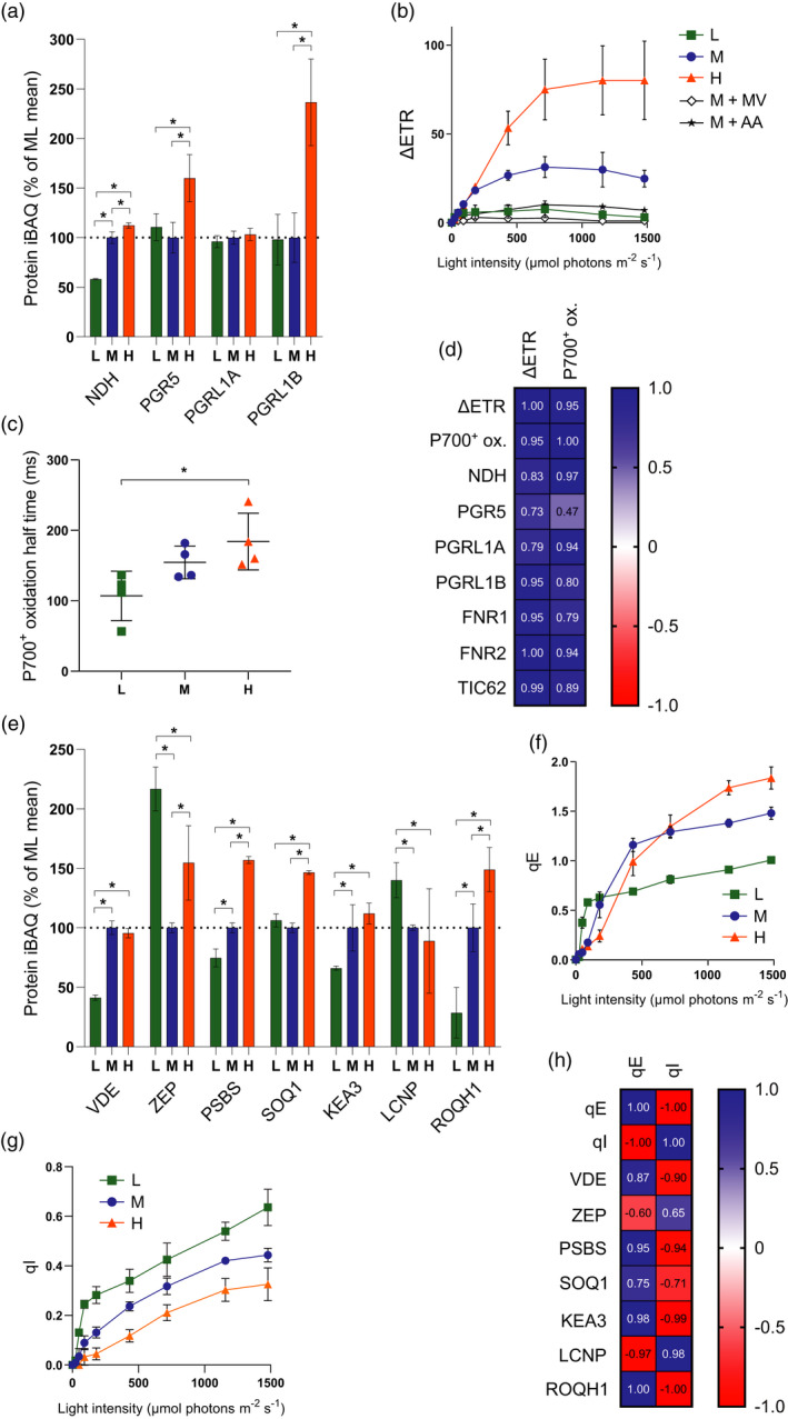
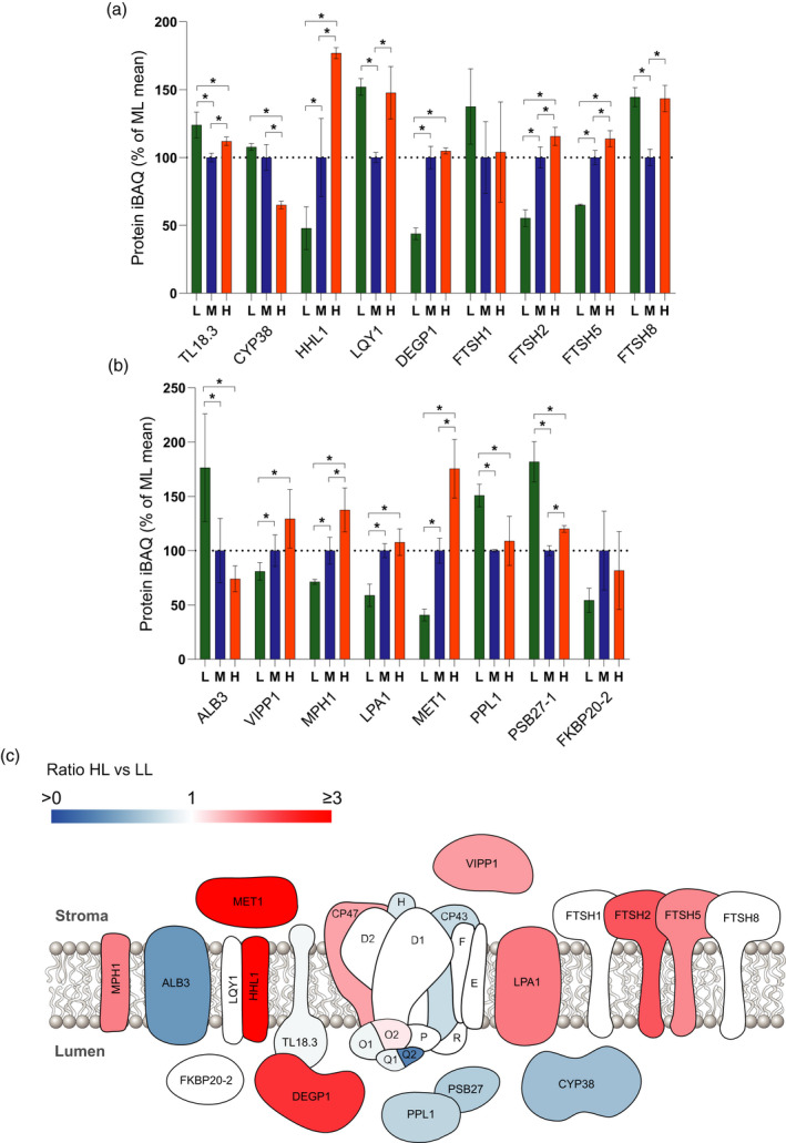
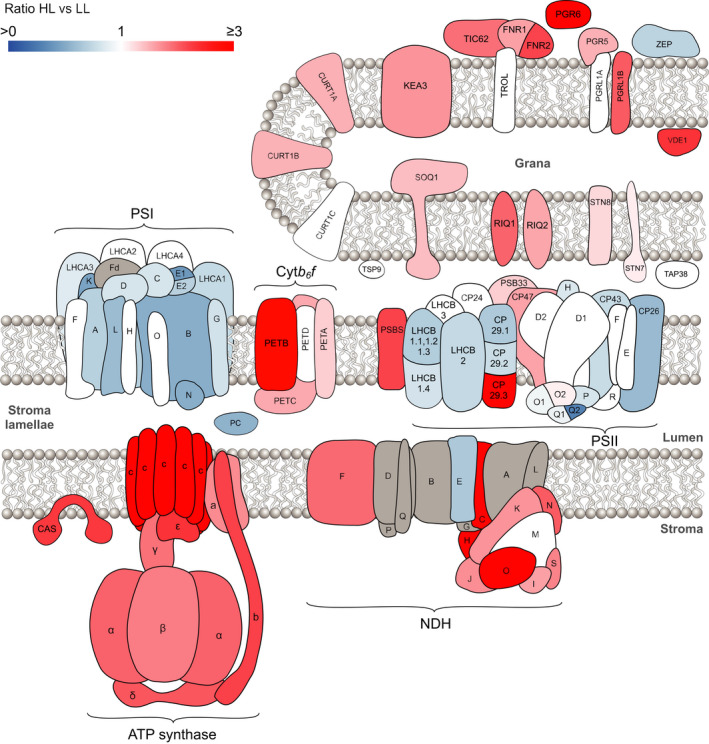
Photosynthetic acclimation, the ability to adjust the composition of the thylakoid membrane to optimise the efficiency of electron transfer to the prevailing light conditions, is crucial to plant fitness in the field. While much is known about photosynthetic acclimation in Arabidopsis, to date there has been no study that combines both quantitative label-free proteomics and photosynthetic analysis by gas exchange, chlorophyll fluorescence and P700 absorption spectroscopy. Using these methods we investigated how the levels of 402 thylakoid proteins, including many regulatory proteins not previously quantified, varied upon long-term (weeks) acclimation of Arabidopsis to low (LL), moderate (ML) and high (HL) growth light intensity and correlated these with key photosynthetic parameters. We show that changes in the relative abundance of cytb6 f, ATP synthase, FNR2, TIC62 and PGR6 positively correlate with changes in estimated PSII electron transfer rate and CO2 assimilation. Improved photosynthetic capacity in HL grown plants is paralleled by increased cyclic electron transport, which positively correlated with NDH, PGRL1, FNR1, FNR2 and TIC62, although not PGR5 abundance. The photoprotective acclimation strategy was also contrasting, with LL plants favouring slowly reversible non-photochemical quenching (qI), which positively correlated with LCNP, while HL plants favoured rapidly reversible quenching (qE), which positively correlated with PSBS. The long-term adjustment of thylakoid membrane grana diameter positively correlated with LHCII levels, while grana stacking negatively correlated with CURT1 and RIQ protein abundance. The data provide insights into how Arabidopsis tunes photosynthetic electron transfer and its regulation during developmental acclimation to light intensity.

Keywords: Acclimation; electron transfer; light harvesting; proteomics; thylakoid.


Conflict of interest statement

The authors have no conflict of interest to declare.

Figures

Figure 1
Figure 1
Arabidopsis plants have different morphologies after acclimation under low, moderate and high light. (a) Plants were initially grown under moderate light (ML, Step 1) at 150 µmol photons m−2 sec−1and then (Step 2) acclimated under low light (LL) at 25 µmol photons m−2 sec−1or high light at 800 µmol photons m−2 sec−1(HL) or maintained under ML conditions (see the Experimental Procedures section). The plants were photographed at 2 weeks post‐Step 1, and post‐Step 2 at 7 (LL), 5 (ML) and 4 (HL) weeks. (b) Ratios of Chl a to Chl b and protein to Chl in isolated thylakoids from LL, ML and HL (n = 3, mean ± SD). (c) Immunodetection of D2 (PSII), PSAA (PSI), PETA (cyt b 6 f) and ATPH (ATP synthase) in Arabidopsis thylakoid membranes acclimated to LL, ML and HL. Sample loading was normalised to chlorophyll. (d) BN‐PAGE of solubilised stromal lamellae (SL) and granal (G) thylakoid fractions. (e) The 77 K fluorescence emission spectra of LL (green), ML (blue) and HL (orange) thylakoids using 435 nm excitation. (f) The 77 K fluorescence excitation spectra of PSII (695 nm) from LL (green), ML (blue) and HL (orange) thylakoids. (g) The 77 K fluorescence excitation spectra of PSI (735 nm) from LL (green), ML (blue) and HL (orange) thylakoids.
Figure 2
Figure 2
Relative quantification of the major photosynthetic complexes by mass spectrometry. (a) MS analysis showing the relative abundance in low (L), moderate (M) and high (H) light‐acclimated thylakoids of key photosynthetic complexes PSII, PSI, LHCII, cyt b 6 f and ATP synthase, expressed as a percentage of the mean at ML. The bars represent the average of three independent peptide preparations (n = 3), derived from a pooled thylakoid sample from 15 plants, which were subject to MS analysis in triplicate in a randomised order and the values were averaged. Data are presented as mean ± SD. Significant differences between light conditions were determined by a modified Welch t‐test (* q < 0.05). (b) MS analysis showing the relative abundance of LHCII trimer and minor monomeric PSII antenna subunits. Sampling details are as stated above. (c) MS analysis showing the abundance of LHCI subunits relative to PSI. Sampling details are as stated above.
Figure 3
Figure 3
Assessment of changes in thylakoid membrane grana size during developmental acclimation. (a) Thin section electron micrographs of chloroplasts in plants acclimated to LL (top row, L), ML (middle row, M) and HL (bottom row, H) (scale bar: 0.5 µm). (b) Number of membrane layers per grana stack calculated from electron microscopy images of chloroplasts in LL (n = 379 grana stacks), ML (n = 354) and HL (n = 507) leaves (one‐way anova with Tukey’s multiple comparisons, ****P < 0.0001). Data are presented as mean ± SD. (c) 3D‐SIM images (shown as Max Projections on the z‐axis with tricubic sharp interpolation) of chloroplasts in plants acclimated to LL (top row, L), ML (middle row, M) and HL (bottom row, H) (scale bar: 0.5 µm). (d) Full‐width at half‐maximum (FWHM) fluorescence intensity of the fluorescent spots (grana) in three‐dimensional SIM images of chloroplasts in LL (n = 97), ML (n = 100) and HL (n = 100) leaves (one‐way anova with Tukey’s multiple comparisons, ****P < 0.0001). Data are presented as mean ± SD. (e) MS analysis showing the relative abundance of proteins involved in the modulation of thylakoid membrane architecture, expressed as a percentage of the mean at ML. Sampling details are as stated in Figure 2. (f) Pearson correlation of the mean number of membrane layers per granum and grana FWHM with protein iBAQ values of LHCII trimers, CURT1A, CURT1B and RIQ1. Blue panels indicate a positive correlation while red panels indicate a negative correlation.
Figure 4
Figure 4
Assessment of changes in linear electron transfer (LET) capacity and CO2 assimilation during developmental acclimation. (a) A CO2 as a function of light intensity. Inset highlights gas exchange under low light and the calculated light compensation point. (b) PSII light use efficiency (ΦPSII) as a function of light intensity. (c) Estimated electron transfer rate of PSII (ETR(II)) as a function of light intensity. (d) The fraction of closed PSII reaction centres (1 − qP). (e) PSI light use efficiency (ΦPSI) as a function of light intensity. (f) PSI donor side limitation (Y (ND)). (g) PSI donor side limitation (Y (NA)). (h) MS analysis showing the relative abundance of electron transfer proteins, expressed as a percentage of the mean at ML. Sampling details are as stated in Figure 2. (i) Pearson correlation of the maximum A CO2 and ETR(II) with protein iBAQ values of PGR6, cytb 6 f, ATP synthase, FNR1, FNR2 and TIC62. Blue panels indicate a positive correlation while red panels indicate a negative correlation. For (a–g), HL = orange, ML = blue, LL = green. n = 4, with the exception of (a), where n = 3. Asterisks denote significance (one‐way anova with Tukey’s multiple comparisons, *P < 0.05). Error bars denote SEM.
Figure 5
Figure 5
Assessment of changes in cyclic electron transfer (CET) and non‐photochemical quenching (NPQ) during developmental acclimation. (a) MS analysis showing the relative abundance of CET‐related proteins, expressed as a percentage of the mean at ML. Sampling details are as stated in Figure 2. (b) Difference in estimated electron transfer rate (ΔETR) between PSI (ETR(I)) and PSII (ETR(II)) versus light intensity. (c) P700 oxidation half‐time upon illumination with 255 μmol photons m−2 sec−1 740 nm light (one‐ way anova with Tukey’s multiple comparisons, *P < 0.05). (d) Pearson correlation of the maximum ΔETR and P700+ oxidation half‐time with protein iBAQ values of NDH, PGR5, PGRL1A, PGRL1B, FNR1, FNR2 and TIC62. (e) MS analysis showing the relative abundance of NPQ‐related proteins, expressed as a percentage of the mean at ML. Sampling details are as stated in Figure 2. (f) Rapidly reversible NPQ (qE) of chlorophyll fluorescence versus light intensity. (g) Slowly reversible NPQ (qI) of chlorophyll fluorescence versus light intensity. Sampling details are as stated in Figure 4 and are the same for (b), (c), (f) and (g). For (b), data for ML leaves infiltrated with methyl viologen and with antimycin A are indicated by M + MV and M + AA, respectively. (h) Pearson correlation of the maximum qE and qI with protein iBAQ values of NPQ‐related proteins.
Figure 6
Figure 6
Changing abundance of the PSII repair cycle machinery upon developmental acclimation. (a,b) MS analysis showing the relative abundance of proteins involved in PSII repair, expressed as a percentage of the mean at ML. Sampling details are as stated in Figure 2. (c) Diagram indicating the abundance of PSII repair proteins in HL versus LL. Blue proteins are more abundant in LL, whereas red/pink proteins are more abundant in HL. For quantified proteins where no significant difference was detected, they are displayed in white.
Figure 7
Figure 7
A comparison of high light versus low light acclimation in the thylakoid membrane proteome. Schematic diagram indicating the relative abundance of thylakoid proteins in HL versus LL. Blue proteins are more abundant in LL, whereas red/pink proteins are more abundant in HL. Where no significant difference was detected for a quantified protein it is displayed in white. Proteins not identified by MS analysis are shown in grey.

References

    1. Adams, W.W. , Watson, A.M. , Mueh, K.E. , Amiard, V. , Turgeon, R. , Ebbert, V. , Logan, B.A. , Combs, A.F. and Demmig‐Adams, B. (2007) Photosynthetic acclimation in the context of structural constraints to carbon export from leaves. Photosynth. Res. 94, 455–466. - PubMed
    1. Albanese, P. , Manfredi, M. , Meneghesso, A. , Marengo, E. , Saracco, G. , Barber, J. , Morosinotto, T. and Pagliano, C. (2016) Dynamic reorganization of photosystem II supercomplexes in response to variations in light intensities. Biochim. Biophys. Acta – Bioenerg. 1857, 1651–1660. - PubMed
    1. Albanese, P. , Manfredi, M. , Re, A. , Marengo, E. , Saracco, G. and Pagliano, C. (2018) Thylakoid proteome modulation in pea plants grown at different irradiances: quantitative proteomic profiling in a non‐model organism aided by transcriptomic data integration. Plant J. 96, 786–800. - PubMed
    1. Albertsson, P.A. , Andreasson, E. , Stefansson, H. and Wollenberger, L. (1994) Fractionation of thylakoid membrane. Methods Enzymol. 228, 469–482.
    1. Amstutz, C.L. , Fristedt, R. , Schultink, A. , Merchant, S.S. , Niyogi, K.K. and Malnoë, A. (2020) An atypical short‐chain dehydrogenase–reductase functions in the relaxation of photoprotective qH in Arabidopsis. Nat. Plants, 6, 154–166. - PMC - PubMed

Publication types