. 2020 Dec;9(1):2361-2367.
doi: 10.1080/22221751.2020.1837017.

A SARS-CoV-2 variant with the 12-bp deletion at E gene

Affiliations

A SARS-CoV-2 variant with the 12-bp deletion at E gene

Yi-Sheng Sun et al. Emerg Microbes Infect. 2020 Dec.

Abstract

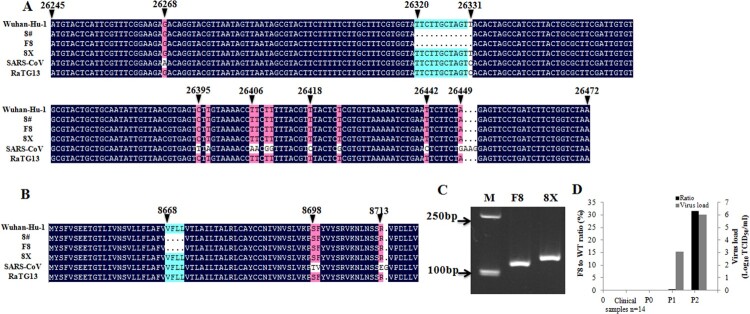
The coronavirus disease 2019 (COVID-19) pandemic is still ongoing and has become an important public health threat. This disease is caused by a new coronavirus named severe acute respiratory syndrome coronavirus-2 (SARS-CoV-2) infection, and so far, little is known about this virus. In this study, by using plaque purification, we purified two SARS-CoV-2 virus strains from the same specimen, one named F8 containing a 12-bp deletion in the E gene and the other named 8X containing the wild-type E gene. There was no significant difference in the viral titer and infectivity of these two strains. The S protein content of the F8 viral culture was 0.39 μg/ml, much higher than that of 8X. An inactivated vaccine made from the F8 strain could trigger high levels of the IgG titer and neutralizing antibody titer, which could last for at least 6 weeks and were significantly higher than those from the 8X strain at 1 and 3 weeks post vaccination, respectively. In conclusion, we reported that both the E gene mutant and wild-type SARS-CoV-2 strains were isolated from the same clinical sample by plaque purification. A 12-bp deletion in the E gene was important for SARS-CoV-2 replication and immunogenicity.

Keywords: COVID-19; E gene mutant; SARS-CoV-2; immunogenicity; virus replication.


Conflict of interest statement

No potential conflict of interest was reported by the author(s).

Figures

Figure 1.
Figure 1.
A 12-bp deletion in the E gene found in SARS-CoV-2. Nucleotide (A) and amino acid (B) sequence alignments of the E genes of SARS-CoV Tor2, bat coronavirus RaTG13, and four SARS-CoV-2 strains (8#, F8, 8X, Wuhan-Hu-1). (C) Electrophoresis of the partial E gene PCR products from F8 and 8X. M: marker. (D) Detection of the E gene mutant in 14 clinical specimens, and in the original specimen and passages 1 and 2 (P0, P1 and P2) of viral culture before plaque purification. Viral loads of P1 and P2 were quantitated by Tagman RT-PCR based on the standard curves obtained from 10-fold serial dilutions of the F8 strain with 106.5 TCID50/ml.
Figure 2.
Figure 2.
Viral titer, S protein content, and infectivity of the F8 and 8X strains. (A) Vero cells were infected with the F8 or 8X virus at an MOI of 0.01 for 48 h. PBS was used as the negative control. Bar: 200 μm. (B) Vero cells were infected with the F8 or 8X virus for 48 h and an indirect immunofluorescence assay using the positive serum from a COVID-19 recovered patient was performed. SARS-CoV-2 virus was stained with the FITC-labeled secondary antibody (Green). Nuclei were stained with DAPI (Blue). Bar: 100 μm. (C) Viral titers (Black) and S protein content (Grey) of the F8 and 8X strains. *Significant difference between the two groups (p < 0.05).
Figure 3.
Figure 3.
Immunogenicity of the F8 and 8X vaccines in mice. Mice were immunized with F8 and 8X vaccines at day 0 and booster at day 7. Aluminum hydroxide at a concentration of 0.5 mg/ml in PBS was used as the negative control. The S protein content was used to quantify the vaccine dose. High dose: 135 ng of S protein. Low dose: 27 ng of S protein. Each group had six mice. Serum was collected at 1, 2, 3, 4, 5, and 6 weeks post booster vaccination. (A) SARS-CoV-2-specific IgG antibody titers were determined by ELISA. *Significant difference between the two groups (p < 0.05). (B) Serum samples at 1, 3, and 6 weeks post the booster vaccination were obtained, and the neutralizing antibody titer was measured by the plaque reduction neutralization test. Serum dilutions leading to 50% plaque reductions (PRNT50) were calculated as titers. *Significant difference between the two groups (p < 0.05).

References

    1. Guan W-j, Ni Z-y, Hu Y, et al. . Clinical characteristics of coronavirus disease 2019 in China. N Engl J Med. 2020 Apr;382(18):1708–1720. - PMC - PubMed
    1. Zhang R, Li Y, Zhang AL, et al. . Identifying airborne transmission as the dominant route for the spread of COVID-19. Proc Natl Acad Sci USA. 2020;117(26):14857–14863. - PMC - PubMed
    1. WHO . WHO coronavirus disease (COVID-19) dashboard. Geneva: World Health Organization; 2020; [cited 2020 Aug 16]. Available from: https://COVID19.who.int/.
    1. Zhu N, Zhang D, Wang W, et al. . A novel coronavirus from patients with pneumonia in China, 2019. N Engl J Med. 2020 Feb;382(8):727–733. - PMC - PubMed
    1. Lu R, Zhao X, Li J, et al. . Genomic characterisation and epidemiology of 2019 novel coronavirus: implications for virus origins and receptor binding. Lancet. 2020 Feb;395(10224):565–574. - PMC - PubMed

MeSH terms

Substances

LinkOut - more resources