

Neurovascular coupling and bilateral connectivity during NREM and REM sleep
- PMID: 33118932
- PMCID: PMC7758068
- DOI: 10.7554/eLife.62071
Neurovascular coupling and bilateral connectivity during NREM and REM sleep
Abstract
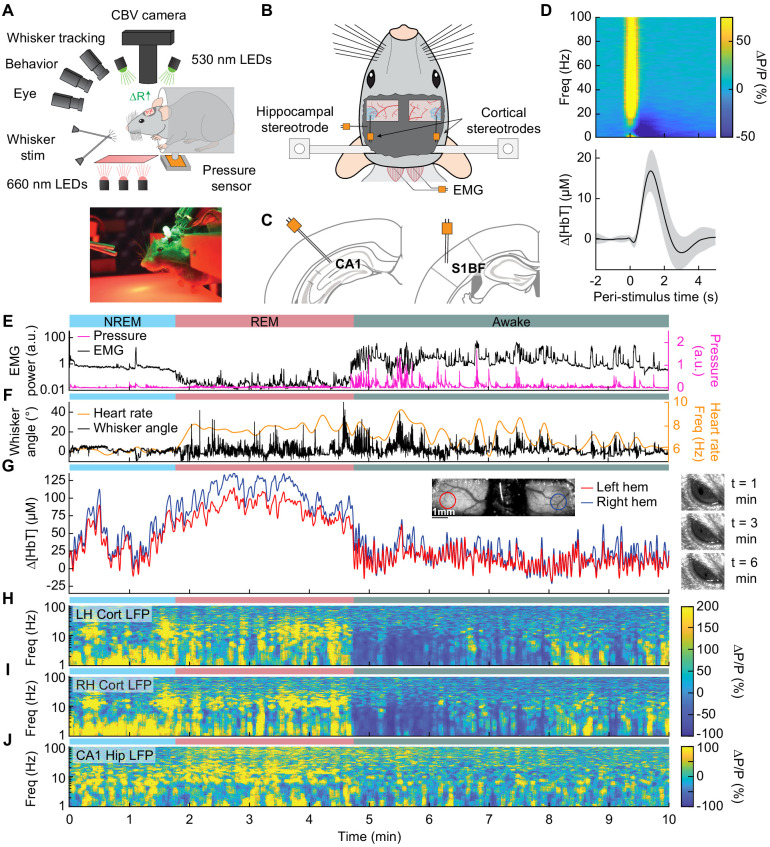
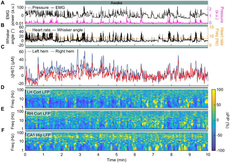
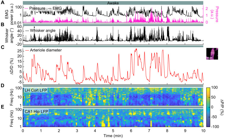
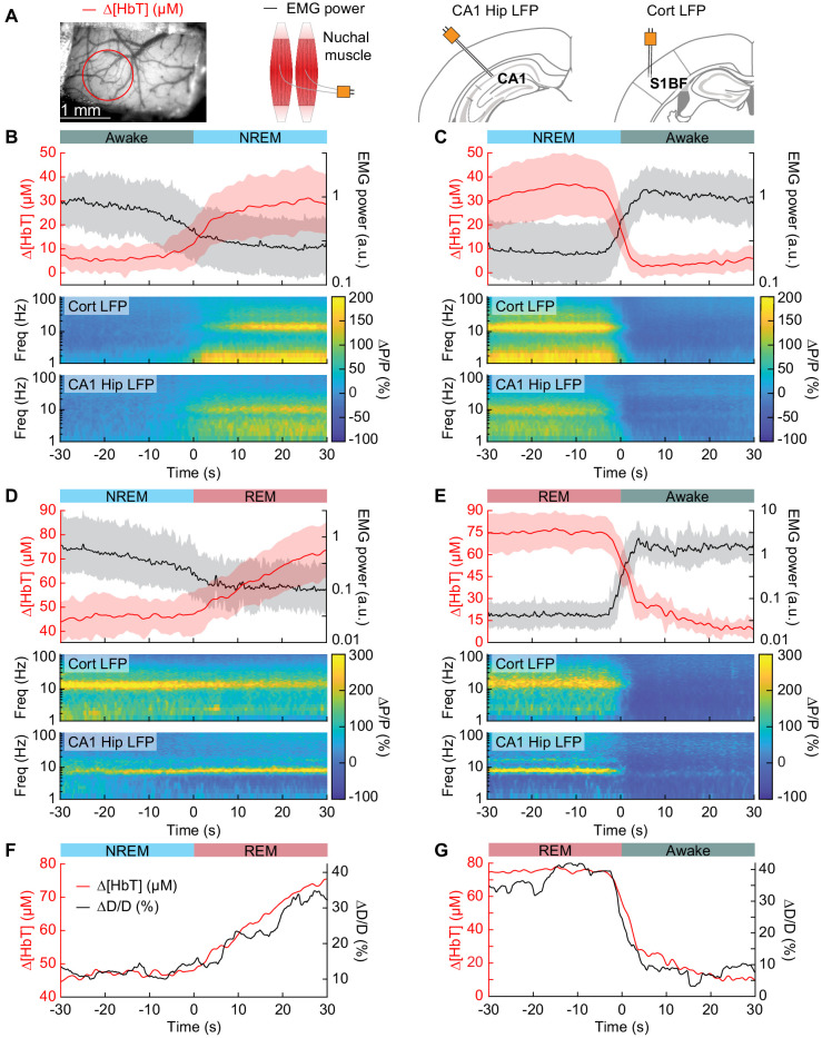
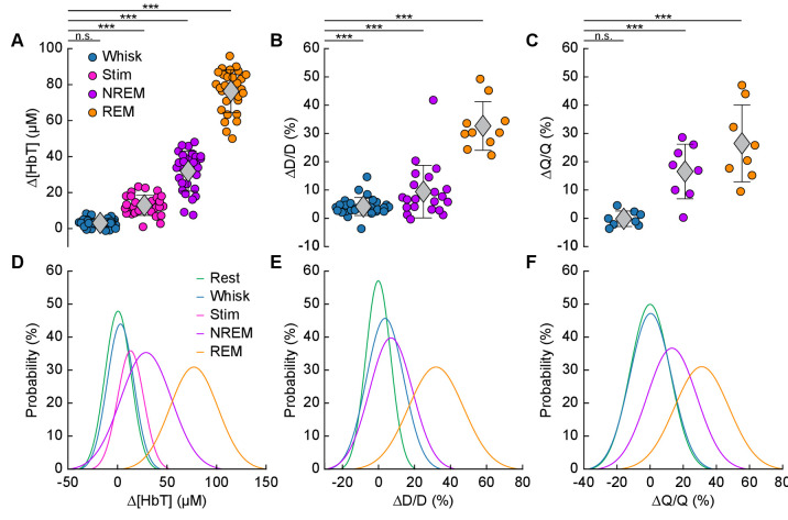
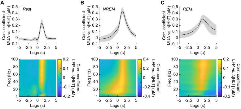
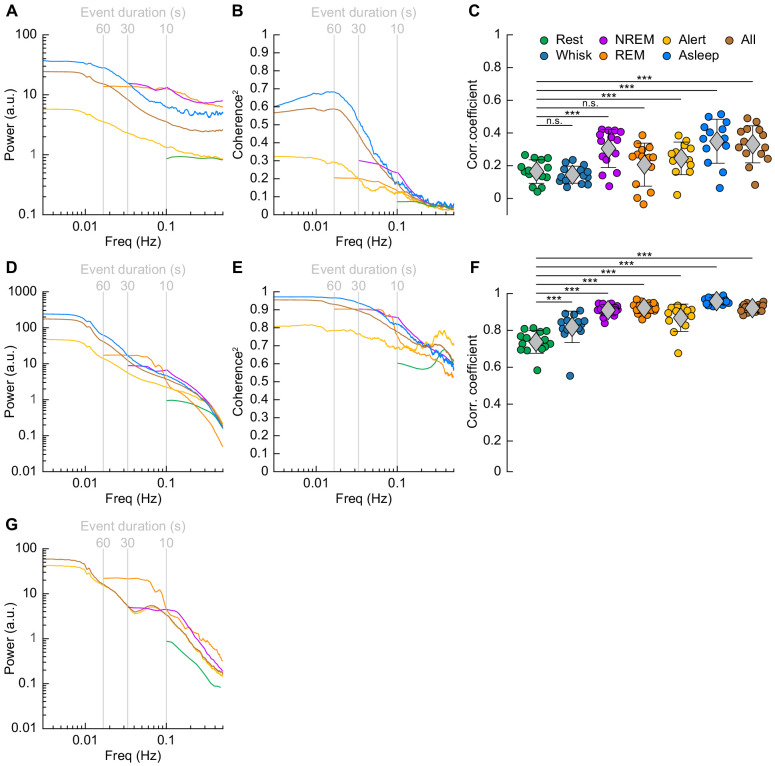
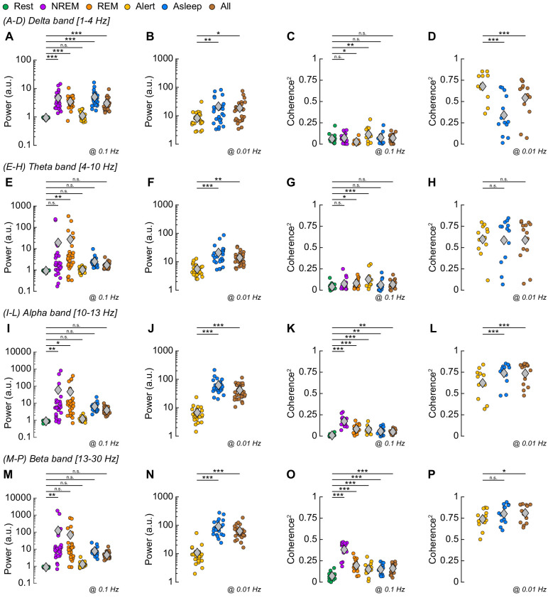
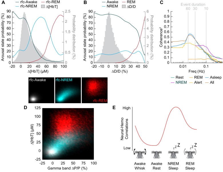
To understand how arousal state impacts cerebral hemodynamics and neurovascular coupling, we monitored neural activity, behavior, and hemodynamic signals in un-anesthetized, head-fixed mice. Mice frequently fell asleep during imaging, and these sleep events were interspersed with periods of wake. During both NREM and REM sleep, mice showed large increases in cerebral blood volume ([HbT]) and arteriole diameter relative to the awake state, two to five times larger than those evoked by sensory stimulation. During NREM, the amplitude of bilateral low-frequency oscillations in [HbT] increased markedly, and coherency between neural activity and hemodynamic signals was higher than the awake resting and REM states. Bilateral correlations in neural activity and [HbT] were highest during NREM, and lowest in the awake state. Hemodynamic signals in the cortex are strongly modulated by arousal state, and changes during sleep are substantially larger than sensory-evoked responses.
Keywords: 2-photon microscopy; arousal state; electrophysiology; mouse; neuroscience; neurovascular coupling; optical imaging; sleep.
© 2020, Turner et al.
Conflict of interest statement
KT, KG, EP, PD No competing interests declared
Figures


Comment in
-
Supplying the sleeping brain.Elife. 2020 Dec 23;9:e64597. doi: 10.7554/eLife.64597. Elife. 2020. PMID: 33355090 Free PMC article.
References
Publication types
MeSH terms
Associated data
Grants and funding
LinkOut - more resources
Full Text Sources
Other Literature Sources
