Review
. 2020 Dec;92(6):e12990.
doi: 10.1111/sji.12990.

Mechanism of fibrosis and stricture formation in Crohn's disease

Affiliations
Review

Mechanism of fibrosis and stricture formation in Crohn's disease

Johannes Alfredsson et al. Scand J Immunol. 2020 Dec.

Abstract

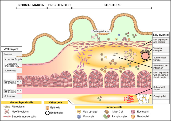
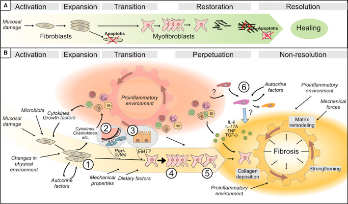
Crohn's disease (CD) is a chronic inflammatory disease of the gastrointestinal tract that leads to substantial suffering for millions of patients. In some patients, the chronic inflammation leads to remodelling of the extracellular matrix and fibrosis. Fibrosis, in combination with expansion of smooth muscle layers, leaves the bowel segment narrowed and stiff resulting in strictures, which often require urgent medical intervention. Although stricture development is associated with inflammation in the affected segment, anti-inflammatory therapies fall far short of treating strictures. At best, current therapies might allow some patients to avoid surgery in a shorter perspective and no anti-fibrotic therapy is yet available. This likely relates to our poor understanding of the mechanism underlying stricture development. Chronic inflammation is a prerequisite, but progression to strictures involves changes in fibroblasts, myofibroblasts and smooth muscle cells in a poorly understood interplay with immune cells and environmental cues. Much of the experimental evidence available is from animal models, cell lines or non-strictured patient tissue. Accordingly, these limitations create the basis for many previously published reviews covering the topic. Although this information has contributed to the understanding of fibrotic mechanisms in general, in the end, data must be validated in strictured tissue from patients. As stricture formation is a serious complication of CD, we endeavoured to summarize findings exclusively performed using strictured tissue from patients. Here, we give an update of the mechanism driving this serious complication in patients, and how the strictured tissue differs from adjacent unaffected tissue and controls.

Keywords: Crohn's disease; fibrosis; strictures.


Conflict of interest statement

Both authors declare no conflict of interest.

Figures

Figure 1
Figure 1
An overview of strictured tissue showing changes in intestinal wall layers and cell populations. A schematic of the most apparent histological changes in the intestinal wall during the progression from normal to strictured tissue is shown. This includes increased intestinal wall thickness due to expanded smooth muscle cells, and hence muscle layers, concomitant with fibrotic changes in the submucosa and subserosa. Histological features described in the text are highlighted in the right column as ‘Key events’ with the location of the event indicated by an arrow. See the text and subsequent figures for details
Figure 2
Figure 2
An overview of mesenchymal cell markers and their distribution in tissue. A, Fibroblasts, myofibroblasts and smooth muscle cells are traditionally distinguished by their staining pattern of the three markers vimentin, α‐SMA and desmin. B, Distribution of the additional markers SMA and FAP in the intestinal walls of strictured (STR) tissue compared to normal (non‐strictured) control (Ctrl) tissue. C, Staining pattern for smooth muscle cells with different phenotypes (based on the degree of differentiation) as described in the text. D, The distribution of smooth muscle cells with different degrees of differentiation in the intestinal wall layers in non‐strictured tissue (Ctrl) and strictured tissue (STR). (A‐C): Green filled circle indicates presence of a marker (positive staining). C,D, Filled circles indicate the presence of a particular SMC phenotype based on the colour coding defined in C. VIM, Vimentin; SMA, alpha‐smooth muscle actin; DES, Desmin; FAP, Fibroblast activating protein; Ctrl, normal non‐strictured tissue; STR, strictured tissue; diff, differentiated; CGA7, antibody that selectively recognizes contractile smooth muscle cells expressing both alpha‐ and gamma‐actin; SMC, Smooth muscle cell. The staining patterns reflect data from numerous publications and mostly reflect immunohistochemical observations
Figure 3
Figure 3
Schematic of the strictured wall with a focus on location of mesenchymal cells. A, The transition into strictured tissue involves an increase in extracellular matrix deposition, fibrosis (indicated by yellow/orange) particularly in the submucosa and subserosa, and expansion of the muscle layers along with infiltration of cells. There is accumulation of predominantly FAP+ myofibroblast in the layers most affected by fibrosis, namely, in ① the submucosa and ② subserosa. However, similar enrichment is also observed in the ③ peri‐cryptal area of the mucosa. Grey boxes show the condition in normal (non‐strictured) tissue with arrows showing the changes that occur inside strictured tissue. The accumulated myofibroblasts might have arisen from ④ pericytes, ⑤ matrix‐resident fibroblasts, or ⑧ epithelial cells (through epithelial‐to‐mesenchymal transition). Also depicted is ⑥ phenotypically changed SMCs along the border between the submucosa and the muscle layers and in the ⑦ fibromuscular infiltration in submucosa. (B‐D) shows the subtypes of the mesenchymal cells depicted in (A), as discussed. See the text for further details
Figure 4
Figure 4
Genes and proteins expressed in strictured tissue compared to normal tissue. A, Shows differences in whole tissue analyses of mRNA and/or protein in strictured ileum (upper panel) and colon (lower panel) compared to non‐strictured tissue. B, Shows differences in intestinal wall layer expression of relevant proteins, mRNAs and miRNAs in the mucosa (upper panel) or below the mucosa (lower panel) of strictured tissue compared non‐strictured tissue. Green indicates expression higher than, and red indicates expression lower than, non‐strictured tissue. Standard text indicates mRNA level; * indicates protein level; ** indicates changes detected at both the protein and mRNA levels. § indicates submucosa only. Abbreviations; miR = micro‐RNA
Figure 5
Figure 5
Schematic illustration of the cells and factors involved in the indicated phases of the stricturing process in intestinal tissue. A, The sequence of events during a simplified and idealized normal healing process. This involves activation and expansion of fibroblasts and subsequent transition of fibroblasts into myofibroblasts, which finalize the restoration of the extracellular matrix. The process occurs during a limited time period, and when the repair process is complete, redundant cells undergo apoptosis (indicated by cells with a red ‘x’) and healing is achieved. B, Under the prevailing conditions in the intestinal wall of a subset of Crohn's patients, the ‘normal’ healing process is aberrant. The pro‐inflammatory environment (red cogwheel) activates and fuels the activation and recruitment of myofibroblast precursors, which include ①matrix‐resident fibroblasts, ② pericytes and ③ epithelial cells. The transition to myofibroblasts can be further facilitated in the presence of certain environmental cues. Released products from activated fibroblasts, as well as from immune cells, create a positive feedback loop so the cogwheel keeps getting fuelled. This results in a shift from an initial inflammation‐driven process to an inflammation‐facilitated process, as the activated mesenchymal cells successively show independence from stimulatory signals. Subsequently, myofibroblasts ④ expand, and ⑤ migrate into the ‘injured’ site where they secrete collagen. Newly formed matrix is strengthened by contraction and cross‐linking, as indicated in the Fibrosis area (yellow cogwheel). However, in a setting where pro‐inflammatory mediators are additionally supplied by immune cells, and perhaps also by ⑥ infiltrating smooth muscle cells, matrix degradation is also stimulated. This leads to a vicious cycle with alternating deposition and degradation of matrix, leading to non‐resolution and fibrosis. Cell symbols are as defined in the Figure 1 and 3 legends

References

    1. Molodecky NA, Soon IS, Rabi DM, et al. Increasing incidence and prevalence of the inflammatory bowel diseases with time, based on systematic review. Gastroenterology. 2012;142(1):46‐54.e42; quiz e30. - PubMed
    1. Torres J, Mehandru S, Colombel J‐F, Peyrin‐Biroulet L. Crohn's disease. Lancet. 2017;389(10080):1741‐1755. - PubMed
    1. Cosnes J, Cattan S, Blain A, et al. Long‐term evolution of disease behavior of Crohn's disease. Inflamm Bowel Dis. 2002;8(4):244‐250. - PubMed
    1. Satsangi J, Silverberg MS, Vermeire S, Colombel JF. The Montreal classification of inflammatory bowel disease: controversies, consensus, and implications. Gut. 2006;55(6):749‐753. - PMC - PubMed
    1. Rieder F, Latella G, Magro F, et al. European Crohn's and colitis organisation topical review on prediction, diagnosis and management of fibrostenosing Crohn's disease. J Crohns Colitis. 2016;10(8):873‐885. - PubMed

Grants and funding