. 2020 Dec 2;48(21):12348-12364.
doi: 10.1093/nar/gkaa909.

Tethering-facilitated DNA 'opening' and complementary roles of β-hairpin motifs in the Rad4/XPC DNA damage sensor protein

Affiliations

Tethering-facilitated DNA 'opening' and complementary roles of β-hairpin motifs in the Rad4/XPC DNA damage sensor protein

Debamita Paul et al. Nucleic Acids Res. .

Abstract

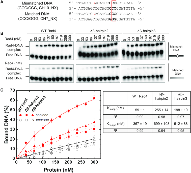
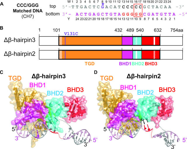
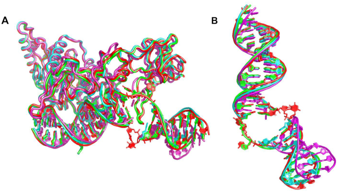
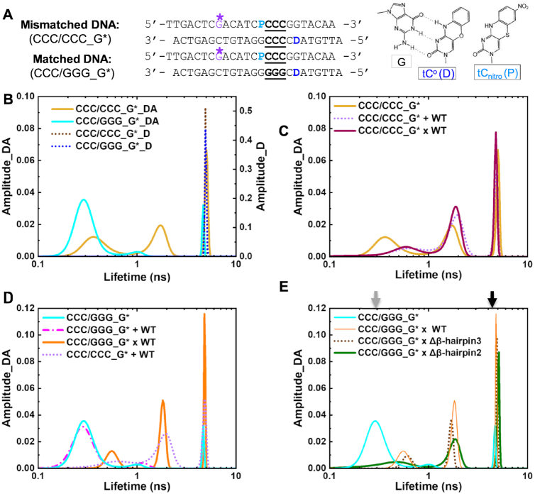
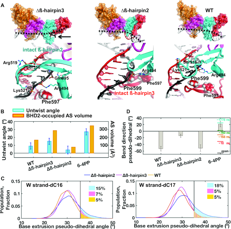
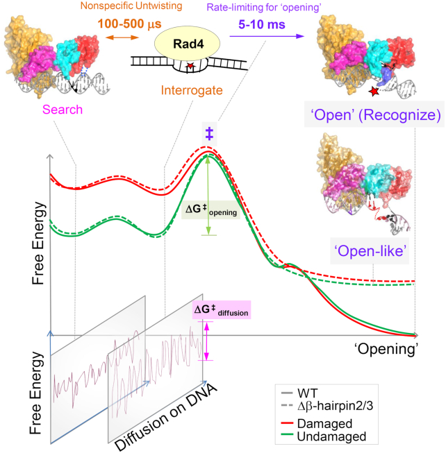
XPC/Rad4 initiates eukaryotic nucleotide excision repair on structurally diverse helix-destabilizing/distorting DNA lesions by selectively 'opening' these sites while rapidly diffusing along undamaged DNA. Previous structural studies showed that Rad4, when tethered to DNA, could also open undamaged DNA, suggesting a 'kinetic gating' mechanism whereby lesion discrimination relied on efficient opening versus diffusion. However, solution studies in support of such a mechanism were lacking and how 'opening' is brought about remained unclear. Here, we present crystal structures and fluorescence-based conformational analyses on tethered complexes, showing that Rad4 can indeed 'open' undamaged DNA in solution and that such 'opening' can largely occur without one or the other of the β-hairpin motifs in the BHD2 or BHD3 domains. Notably, the Rad4-bound 'open' DNA adopts multiple conformations in solution notwithstanding the DNA's original structure or the β-hairpins. Molecular dynamics simulations reveal compensatory roles of the β-hairpins, which may render robustness in dealing with and opening diverse lesions. Our study showcases how fluorescence-based studies can be used to obtain information complementary to ensemble structural studies. The tethering-facilitated DNA 'opening' of undamaged sites and the dynamic nature of 'open' DNA may shed light on how the protein functions within and beyond nucleotide excision repair in cells.


Figures

Figure 1.
Figure 1.
Characterization of DNA binding affinities by Rad4–Rad23 complexes. (A) Sequences of DNA constructs used for the competitive EMSA. The position of the two flipped-out nucleotide pairs in the ‘open’ conformation is indicated with a red box. (B) Typical gel images showing the various Rad4 constructs binding to the mismatched CCC/CCC (top) and matched CCC/GGG DNA (bottom). (C) Quantification of the percent bound DNA fractions in (B) versus protein concentrations. The symbols and error bars indicate the means and ranges as calculated by ± sample standard deviation, respectively, from triplicate gel shift experiments. Filled red symbols indicate mismatched DNA and empty black ones indicate matched DNA; circle, square and triangle indicate WT, Δβ-hairpin2 and 3, respectively. Lines indicate the fit curves of the data points. Apparent dissociation constants for specific binding to mismatched DNA (Ks,app) and nonspecific binding to matched DNA (Kns,app) are also shown.
Figure 2.
Figure 2.
Overall structures of the β-hairpin deletion mutants of Rad4–Rad23 complexes tethered to CCC/GGG matched DNA. (A) The crosslinkable DNA used for crystallization. The ‘top’ strand (W) is colored in gray and the ‘bottom’ strand (Y) in light pink. The stretch of C/G’s within the sequence is in black and red. The disulfide modified G* is in purple with its chemical structure indicated in the inset. The position of the two flipped-out nucleotide pairs in the ‘open’ conformation is indicated with a red box. (B) The domain arrangements and boundaries of Rad4 used in this study. The transglutaminase domain (TGD) of Rad4 is indicated in orange, β-hairpin domain 1 (BHD1) magenta, BHD2 cyan and BHD3 red. Deleted β-hairpin3 (residues 599–605) in Δβ-hairpin3 and β-hairpin2 (residues 515–527) in Δβ-hairpin2 are indicated in white. Disordered regions in crystals are checkered. The V131C point mutation introduced for disulfide crosslinking is shown in purple. The Rad23 construct was the same as in ref. (24). (C and D) The overall crystal structures of Δβ-hairpin3–DNA (PDB ID: 6UBF) and Δβ-hairpin2–DNA (PDB ID: 6UIN). The color codes are the same as in (A) and (B).
Figure 3.
Figure 3.
Comparison of the ‘open’ and ‘open-like’ crystal structures of Rad4–Rad23–DNA complexes. (A) Superposition of Rad4–Rad23–DNA crystal structures. The ‘open’ structures of WT Rad4 bound to 6–4PP (PDB ID: 6CFI) and of WT Rad4 tethered to CCC/GGG matched DNA (4YIR) are in red and green, respectively. The Δβ-hairpin3 and Δβ-hairpin2 mutants tethered to CCC/GGG matched DNA (cyan and magenta, respectively) show similar ‘open-like’ conformations. (B) Superposition of the DNA molecules extracted from the structures shown in (A). The difference in the DNA tails shown by the Δβ-hairpin2 (magenta) is partly due to the differences in crystal packing (Supplementary Discussion and Supplementary Figure S2).
Figure 4.
Figure 4.
DNA conformational landscapes in solution obtained by FLT measurements. (A) DNA constructs for FLT studies. ‘D’ indicates tCo (FRET donor) and ‘P’ is tCnitro (FRET acceptor). G* is disulfide-modified guanine for tethering. Positions of the 3-bp CCC/CCC mismatches or CCC/GGG matched sequences are underlined. (right) Chemical structures of tCo and tCnitro and Watson–Crick type base pairing of tCo with a guanine (G). (BE) Representative FLT distributions from MEM analyses for different DNA and protein–DNA complexes. ‘_D’ indicate DNA with donor only; ‘_DA’ indicate DNA with donor/acceptor pair. Results in (C–E) are all from DNA_DA. (B) FLT distributions of mismatched CCC/CCC_G* _DA (yellow) or _D (dotted brown) and CCC/GGG_G*_DA (cyan) or _D (dotted blue) (C) FLT distributions of mismatched CCC/CCC_G* DNA when by itself (yellow), non-covalently bound to (‘+’; dotted violet) or site-specifically tethered with (‘x’; deep purple) WT Rad4. (D) FLT distributions of matched CCC/GGG_G* DNA when by itself (cyan), non-covalently bound to (‘+’; dotted magenta) or tethered with (‘x’; orange) WT Rad4. The FLT distribution of CCC/CCC_G* + WT Rad4 is also shown in dotted violet. (E) FLT distributions of CCC/GGG_G* when by itself (cyan) or tethered to WT Rad4 (orange), Δβ-hairpin3 (dotted brown) and Δβ-hairpin2 (green). All amplitudes indicate the normalized, fractional amplitudes. The arrows indicate the lifetimes corresponding to the computed FRET efficiencies for B-DNA conformation (gray) and for the DNA conformation in the Rad4-bound ‘open’ crystal structure (black). Reproducibility of FLT distributions for each sample is shown in Supplementary Figure S6. Full reports of the lifetimes, fractional amplitudes, FRET efficiencies of each peak as well as the sample's average FRET efficiencies are in Supplementary Table S2.
Figure 5.
Figure 5.
Initial binding states of the β-hairpin mutants with the CCC/GGG duplex obtained by MD simulations. (A) (Top) Best representative structures of the initial binding states (IBS) show different effects of β-hairpin truncations. Black dashed lines show DNA bend directions (also see panel D); the minor groove around the GGG sequence (red) is below the dashed lines. (Bottom) Enlarged views from the minor groove side. For Δβ-hairpin3, the intact β-hairpin2 inserts further into the minor groove than in the WT to promote untwisting. For Δβ-hairpin2, the intact β-hairpin3 approaches the potential flipping bases from the major groove more than in the WT. Supplementary Movies S1 and S2 also display major groove views, which provide a good view of the BHD3 hairpin on that side. (B) Untwist angle and BHD2-occupied minor groove AS volume (48) show that untwisting correlates with extent of BHD2’s minor groove occupancy, both of which were more pronounced for Δβ-hairpin3 compared to Δβ-hairpin2. (C) Δβ-hairpin2 promotes extrusion of partner strand bases compared to WT and Δβ-hairpin3. The extrusions are facilitated by Phe599 in the β-hairpin3 that moved closer to DNA than in WT. (D) DNA bend directions show bending toward the minor groove (negative values) for CCC/GGG DNA, unlike with 6–4PP. The values of the bend directions for the 6–4PP along the MD trajectory (at different simulation times) are shown in color dashed lines and the value for the crystal structure of the open complex is in black dashed line (25). The standard deviations of block averaged means (80,81) for the untwist angles (B) and the bend direction pseudo-dihedral angles (D) are shown. Full details of the block averaging method are given in Supplementary Methods.
Figure 6.
Figure 6.
Proposed DNA ‘opening’ trajectory and ‘kinetic gating’ mechanism of Rad4/XPC. The top panel illustrates distinct binding modes for Rad4/XPC as it searches for, interrogates, and recognizes a damaged site, and the time scales for fluctuations between these modes, based on prior studies (18–20). The middle panel shows a schematic free-energy profile along the ‘opening’ trajectory. The faster 100- to 500-μs nonspecific untwisting step entails a smaller energetic barrier than the slower 5- to 10-ms rate-limiting step (‡) of the ‘opening’ process. The rate-limiting step involves sufficiently unwound and bent DNA but with the nucleotides not yet fully or stably flipped out into the BHD2/BHD3 groove (19). The free energy barrier (ΔGopening) for ‘opening’ damaged DNA (red) is naturally lower than that for undamaged DNA (green) as DNA damage destabilizes the B-DNA structure. For Rad4 mutants that are lacking either β-hairpin2 or β-hairpin3, the protein can still overcome ΔGopening (although the barrier could be higher) to form ‘open-like’ structures that exhibit the same extent of unwinding as the fully ‘open’ structure with the WT (18), as monitored by the tCo-tCnitro FRET probes; however, the ‘open-like’ structures with the mutants are less stable and do not show well-resolved flipped-out nucleotides in the crystal structures (this study). MD simulations indicate that there can be more than one pathway that leads to ‘open-like’ or ‘open’ structures and demonstrate that the two β-hairpins function in a concerted manner to promote ‘opening’, but can also compensate for each other when one β-hairpin is lacking. The bottom panel illustrates that for each step along the ‘opening’ trajectory, there is also a kinetically competing process of diffusion of Rad4/XPC along the DNA, characterized by ΔGdiffusion. For undamaged DNA, the high ΔGopening compared with ΔGdiffusion favors the protein diffusing away before ‘opening’ a given site, while for damaged DNA this competition favors ‘opening’. However, when tethered, the diffusion of the protein is blocked and it can ‘open’ that site as long as the ΔGopening is thermally surmountable, suggesting that stalling of Rad4/XPC by another protein may be a mechanism employed by NER to enable the ‘opening’ of more resistant NER lesions.

References

    1. Spivak G. Nucleotide excision repair in humans. DNA Repair (Amst.). 2015; 36:13–18. - PMC - PubMed
    1. Scharer O.D. Nucleotide excision repair in eukaryotes. Cold Spring Harb. Perspect. Biol. 2013; 5:a012609. - PMC - PubMed
    1. Hoogstraten D., Bergink S., Ng J.M., Verbiest V.H., Luijsterburg M.S., Geverts B., Raams A., Dinant C., Hoeijmakers J.H., Vermeulen W. et al. .. Versatile DNA damage detection by the global genome nucleotide excision repair protein XPC. J. Cell Sci. 2008; 121:2850–2859. - PubMed
    1. Sugasawa K., Ng J.M., Masutani C., Iwai S., van der Spek P.J., Eker A.P., Hanaoka F., Bootsma D., Hoeijmakers J.H.. Xeroderma pigmentosum group C protein complex is the initiator of global genome nucleotide excision repair. Mol. Cell. 1998; 2:223–232. - PubMed
    1. Wood R.D. DNA damage recognition during nucleotide excision repair in mammalian cells. Biochimie. 1999; 81:39–44. - PubMed

Publication types

MeSH terms