. 2020 Nov 18;48(20):11536-11550.
doi: 10.1093/nar/gkaa934.

Discovery and development of novel DNA-PK inhibitors by targeting the unique Ku-DNA interaction

Affiliations

Discovery and development of novel DNA-PK inhibitors by targeting the unique Ku-DNA interaction

Navnath S Gavande et al. Nucleic Acids Res. .

Abstract

DNA-dependent protein kinase (DNA-PK) plays a critical role in the non-homologous end joining (NHEJ) repair pathway and the DNA damage response (DDR). DNA-PK has therefore been pursued for the development of anti-cancer therapeutics in combination with ionizing radiation (IR). We report the discovery of a new class of DNA-PK inhibitors that act via a novel mechanism of action, inhibition of the Ku-DNA interaction. We have developed a series of highly potent and specific Ku-DNA binding inhibitors (Ku-DBi's) that block the Ku-DNA interaction and inhibit DNA-PK kinase activity. Ku-DBi's directly interact with the Ku and inhibit in vitro NHEJ, cellular NHEJ, and potentiate the cellular activity of radiomimetic agents and IR. Analysis of Ku-null cells demonstrates that Ku-DBi's cellular activity is a direct result of Ku inhibition, as Ku-null cells are insensitive to Ku-DBi's. The utility of Ku-DBi's was also revealed in a CRISPR gene-editing model where we demonstrate that the efficiency of gene insertion events was increased in cells pre-treated with Ku-DBi's, consistent with inhibition of NHEJ and activation of homologous recombination to facilitate gene insertion. These data demonstrate the discovery and application of new series of compounds that modulate DNA repair pathways via a unique mechanism of action.


Figures

Figure 1.
Figure 1.
(A) Structure of X80 with Ku EMSA and DNA-PK IC50 values.(B) Binding pocket of X80 in the Ku70/80 core. Ku70 is depicted in cyan and Ku80 in magenta. The DNA ring structure is depicted in orange (circular stick model) and X80 compound in red (spheres). (C) Schematic representation of SAR exploration rationale: a pocket surrounding Ring A of X80 compound for further optimization (Ku70 shown as yellow and Ku80 as gray surface).
Figure 2.
Figure 2.
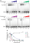
EMSA analysis of Ku inhibitors. (A) Inhibition of Ku–DNA binding as assessed by EMSA. Binding assays were performed as described in ‘Materials and Methods’ section. The indicated compounds were incubated in reaction with DNA and purified Ku, reaction products were separated by native gel electrophoresis. Gels were visualized and quantified by PhosphorImager analysis. (B) Quantification of EMSA data. IC50 values were calculated from a minimum of three independent experiments using GraphPad Prism and data are presented in Table 1.
Figure 3.
Figure 3.
Molecular docking studies (PDB: 1JEQ and 1JEY). (A andB) Molecular interactions of compound 149 (A) and 245 (B) (all in yellow carbon) with Ku70/80 heterodimer (key amino acids are shown in cyan carbon (Ku70), green carbon (Ku80) and cartoon is shown in cyan color). The DNA ring structure is depicted in light orange dots. Interaction with amino acid side chains is indicated with the dashed magenta lines and π–π stacking interactions are shown in solid magenta dumbell. Interaction distances indicated in Å.
Figure 4.
Figure 4.
DNA-PK activity is inhibited by Ku targeted agents. (A and B). Kinases assays were performed as described in ‘Materials and Methods’ section with the indicated compounds. Data are the average and standard error of 2–3 replicates and IC50 values were calculated using GraphPad Prism and data presented in Table 1. (C) Autophosphorylation of DNA-PKcs at the Ser 2056 cluster was assessed in vitro and detected by western blot analysis as described in ‘Materials and Methods’ section. The negative control contained no DNA and ATP and positive control was with DMSO (vehicle). The individual Ku-DBi’s (20 μM), positive inhibitory control (Nu7441, 5 nM) and a negative control (329, 10 μM) were included as indicated. Reaction products were separated by SDS-PAGE, transferred to PVDF and probed sequentially to detect phospho-DNA-PKcs (upper panel) and total DNA-PKcs (lower panel) as indicated.
Figure 5.
Figure 5.
Thermal-shift assay (TSA) of Ku protein. (A) Purified Ku was treated with vehicle or 245 and thermal stability measured by Ku70 western blot. (B) Band intensity was normalized to the 40°C point and plotted versus temperature. Data were fit to a 4-parameter sigmoidal curve and T1/2 determined. The error associated with the fits were <0.5°C. (C) Kinase specificity. The indicated compounds were assessed in a kinase screen using ADP-Glo as described in ‘Materials and Methods’ section. The percent activity remaining for each kinase was calculated and presented as a heat map.
Figure 6.
Figure 6.
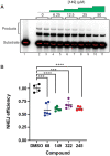
Analysis of Ku targeted DNA-PK inhibitors on in vitro and cellular NHEJ. (A) A radiolabeled linearized plasmid DNA (3 kbp) substrate (lane 1) and the formation of plasmid multimers when increasing concentration of compound 149 was incubated with the extract (lanes 2–11). Quantification of the data revealed an IC50 of ∼15 μM for compound 149. (B) Cellular NHEJ was assessed by host cell reactivation as described in ‘Materials and Methods’ section. Individual data points are presented along with the average and standard error (**** p<-0.0001, *** p<0.001+.
Figure 7.
Figure 7.
Sensitization to DSB inducing agents by Ku targeted DNA-PK inhibitors. (A) Schematic representation of Bleomycin treatment and 245 pre-incubation times used in this assay. H460 cells were incubated for 3, 2 or 1 h with 245 inhibitor (indicated in black arrows) and 1-h Bleomycin treatment was applied to each condition (blue line) prior to fixing the cells. (B) Immunofluorescence staining of DNA-PKcs p(S2056) (green) and γH2A.X (pS139) (red) in H460 cells following treatment. Untreated control cells were incubated with 1% DMSO. Cells were fixed, immunostained and fluorescence microscopy images were digitally captured using 60×-immersion oil magnification and colored; scale bar: 10 μm. (C) Quantification of integrated fluorescence intensity of DNA-PKcs p(S2056). (D) Quantification of γH2AX foci. For (C) and (D) fluorescence microscopy images of H460 cells subjected to treatments described in (A) were digitally captured using 10× magnification and analyzed with the extended version Fiji from ImageJ. Data obtained from 200 cells (black circles) are shown as median (red bar). Statistical analysis was determined using unpaired t-test; ****P< 0.0001; ***P< 0.001. (E) Cellular inhibition of Ku–DNA interaction by compound 68. MEF cells were seeded and treated with vehicle or 20 μM 68 and the indicated Bleomycin concentration. Cell viability was assessed by CCK-8 metabolic assay and data are presented as the mean and SEM of triplicate determinations. (F) Enhancement of cell killing after irradiation. After irradiation, cells were plated to assess clonogenic survival as described and data represent the mean and SEM of triplicate determinations. The * denotes points that differ significantly from the DMSO control with a P < 0.05. (G) Sensitization to etoposide by compound 322. H460 cells were plated and treated with vehicle or 20 μM 322 and the indicated concentration of etoposide for 48 h after which cell viability was determined by CCK-8. Data are presented as the mean and SEM of triplicate determinations.
Figure 8.
Figure 8.
Enhancement of HDR mediated gene insertion. (A) Schematic of gene insertion experiment. The 5.2 kbp donor plasmid was designed with a 2.2 kb insert to express GFP and confer puromycin resistance. Homology arms of 800 bp were included on either end to target the insertion to the genomic EMX1 site. PCR primers were designed to allow amplification of the left and right junctions to assess accurate gene insertion (B) Genomic DNA was analyzed for precise gene insertion by in/out PCR analysis using the primers depicted in panel (A).

References

    1. Abraham R.T. PI 3-kinase related kinases: ‘big’ players in stress-induced signaling pathways. DNA Repair (Amst.). 2004; 3:883–887. - PubMed
    1. Mohiuddin I.S., Kang M.H.. DNA-PK as an emerging therapeutic target in cancer. Front Oncol. 2019; 9:635. - PMC - PubMed
    1. Lieber M.R. The mechanism of double-strand DNA break repair by the nonhomologous DNA end-joining pathway. Annu. Rev. Biochem. 2010; 79:1–31. - PMC - PubMed
    1. Wang Y., Ghosh G., Hendrickson E.A.. Ku86 represses lethal telomere deletion events in human somatic cells. Proc. Natl. Acad. Sci. U.S.A. 2009; 106:12430–12435. - PMC - PubMed
    1. Ruis B.L., Fattah K.R., Hendrickson E.A.. The catalytic subunit of DNA-dependent protein kinase regulates proliferation, telomere length, and genomic stability in human somatic cells. Mol. Cell Biol. 2008; 28:6182–6195. - PMC - PubMed

Publication types