

doi: 10.1007/s00508-020-01753-3.
Clinical-Pathological Conference Series from the Medical University of Graz : Case No 170: A 33-year-old psychologist with severe dyspnea and right-sided chylothorax
Affiliations
- PMID: 33119872
- PMCID: PMC7840639
- DOI: 10.1007/s00508-020-01753-3
Item in Clipboard
Clinical-Pathological Conference Series from the Medical University of Graz : Case No 170: A 33-year-old psychologist with severe dyspnea and right-sided chylothorax
Wien Klin Wochenschr.
2021 Jan.
No abstract available
Keywords: Cystic lung disease; Lymphangioleiomyomatosis; Sirolimus; VEGF-D.
Conflict of interest statement
P.K. Bauer, M. Flicker, E. Fabian, H. Flick, L. Brcic, B. Liegl-Atzwanger, M. Janisch, M. Fuchsjäger, H. Olschewski and G.J. Krejs declare that they have no competing interests.
Figures

Chest X‑ray (in expiration) with subtotal opacity of the right lung and merely residual ventilation of the right upper lobe (arrow) as well as a mediastinal shift to the left
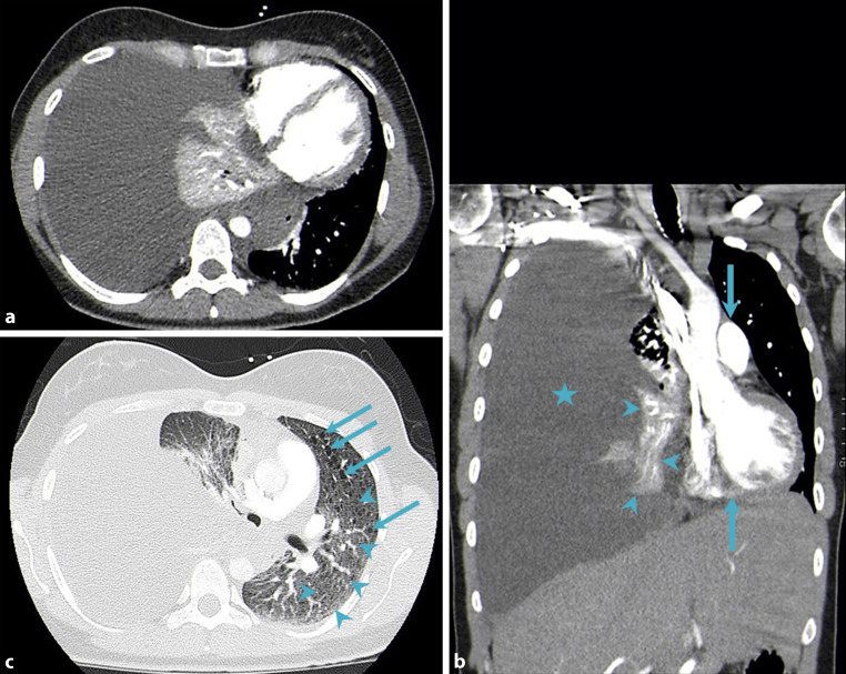
CT of the chest. Axial (a) and coronal (b) images (soft tissue window setting); massive right pleural effusion (asterisk), atelectasis of the right middle and lower lung lobes (arrowheads) as well as a marked mediastinal shift to the left (arrows). Axial image (c; lung window setting); multiple small lung cysts (diameter up to 6 mm; arrows) as well as extensive ground glass opacities (arrowheads)
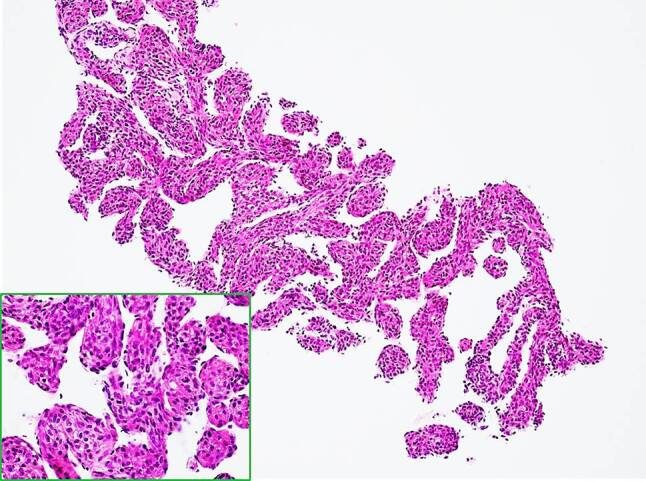
Histological presentation of the retroperitoneal mass. Spindle tumor cells without marked atypia are present, displaying eosinophilic cytoplasm and interspersed blood vessel structures (hematoxylin-eosin staining, objective ×4 and ×20 inlet)
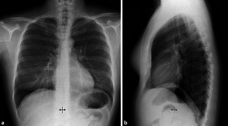
Normal follow-up chest X‑ray a; p.a. and b; lateral) after treatment with sirolimus for 6 months
References
-
- Light RW. Clinical practice. Pleural effusion. N Engl J Med. 2002;346(25):1971–1977. - PubMed
-
- Doerr CH, Allen MS, Nichols FC, Ryu JH. Etiology of chylothorax in 203 patients. Mayo Clin Proc. 2005;80(7):867–870. - PubMed
-
- Agrawal V, Doelken P, Sahn SA. Pleural fluid analysis in chylous pleural effusion. Chest. 2008;133(6):1436–1441. - PubMed
-
- Braun SR, Everett ED, Perry MC, Sunderrajan EV. Concise textbook of pulmonary medicine. New York: Elsevier; 1989.
MeSH terms
Substances
LinkOut - more resources
Full Text Sources
Medical
