

Case Reports
doi: 10.4103/ijo.IJO_2575_20.
Central retinal vein occlusion with COVID-19 infection as the presumptive etiology
Affiliations
- PMID: 33120696
- PMCID: PMC7774137
- DOI: 10.4103/ijo.IJO_2575_20
Item in Clipboard
Case Reports
Central retinal vein occlusion with COVID-19 infection as the presumptive etiology
Indian J Ophthalmol.
2020 Nov.
Abstract
Thromboembolic phenomenon related to Coronavirus disease 2019 (COVID-19) has been well documented in literature; however, reported ocular manifestations of COVID-19 are limited to vision sparing ocular conditions like conjunctivitis. We report a case of a 17-year-old female who presented to us with central retinal vein occlusion with proven recent past COVID-19 infection as presumed etiology which was not known to her at the time of presentation.
Keywords: Central retinal vein occlusion; coronavirus; covid19; ocular manifestations of covid-19.
Conflict of interest statement
None
Figures
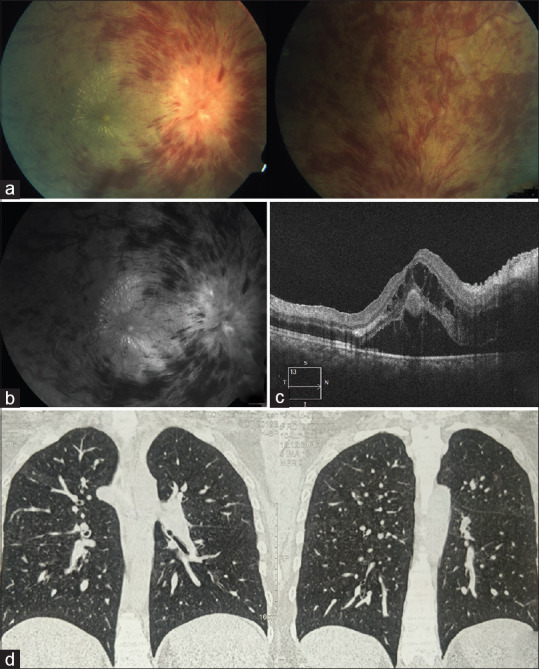
(a) Color and (b) red-free fundus images of OD showing optic disc edema and multiple hemorrhages in all quadrants with a macular star. (c) Optical coherence tomography scan of macula shows neurosensory detachment and cystoid macular edema. (d) Computed tomography scan of the chest showing multiple ground glass opacities

(a) Color fundus photo of the right eye showing resolution of signs after the first injection on the left and further resolution after the second injection in the image on the right. (b) Disc photo shows resolving disc edema after the first injection on the left side and resolved peripheral retinal hemorrhages after the second injection in the right-side image. (c) OCT of macula of the right eye shows dramatic resolution of CME after the first injection in the left-side image and near total resolution of CME with mild RPE atrophy and ellipsoid layer thinning noted in the right-side image
References
Publication types
MeSH terms
LinkOut - more resources
Full Text Sources
Miscellaneous
