

Synthesis, Physicochemical Characterization, and Cytotoxicity Assessment of Rh Nanoparticles with Different Morphologies-as Potential XFCT Nanoprobes
- PMID: 33120889
- PMCID: PMC7692549
- DOI: 10.3390/nano10112129
Synthesis, Physicochemical Characterization, and Cytotoxicity Assessment of Rh Nanoparticles with Different Morphologies-as Potential XFCT Nanoprobes
Abstract
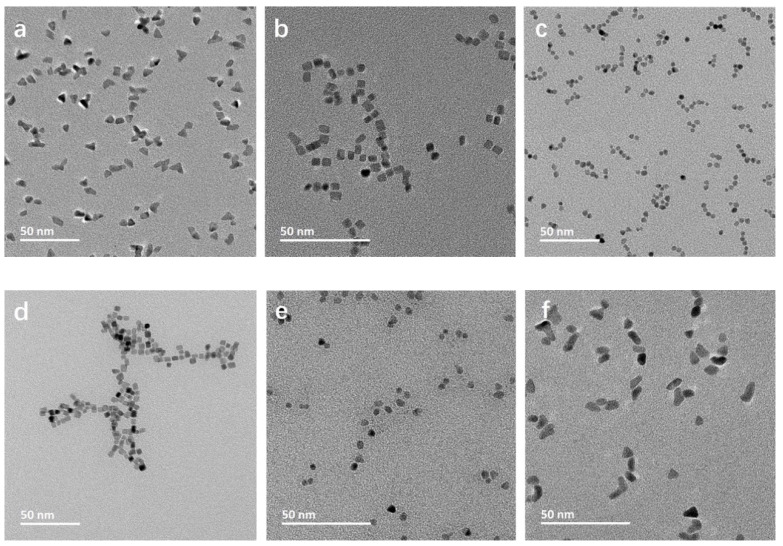
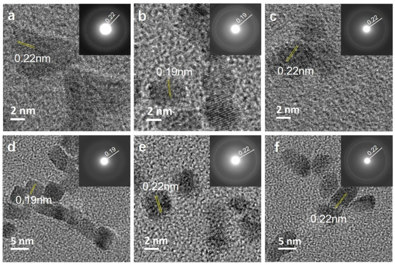
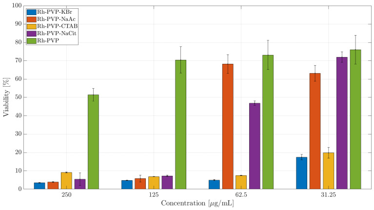
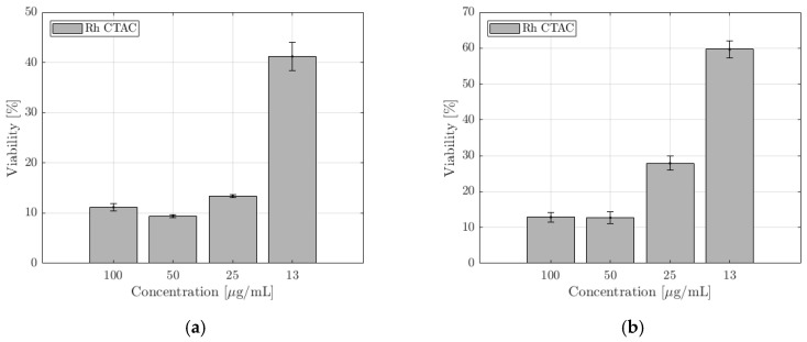
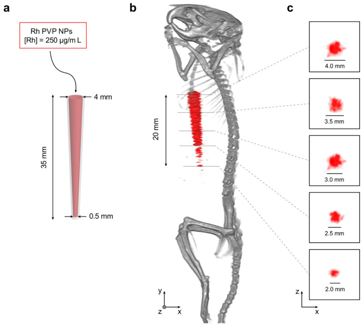
Morphologically controllable synthesis of Rh nanoparticles (NPs) was achieved by the use of additives during polyol synthesis. The effect of salts and surfactant additives including PVP, sodium acetate, sodium citrate, CTAB, CTAC, and potassium bromide on Rh NPs morphology was investigated. When PVP was used as the only additive, trigonal NPs were obtained. Additives containing Br- ions (CTAB and KBr) resulted in NPs with a cubic morphology, while those with carboxyl groups (sodium citrate and acetate) formed spheroid NPs. The use of Cl- ions (CTAC) resulted in a mixture of polygon morphologies. Cytotoxicity of these NPs was evaluated on macrophages and ovarian cancer cell lines. Membrane integrity and cellular activity are both influenced to a similar extent, for both the cell lines, with respect to the morphology of Rh NPs. The cells exposed to trigonal Rh NPs showed the highest viability, among the NP series. Particles with a mixed polygon morphology had the highest cytotoxic impact, followed by cubic and spherical NPs. The Rh NPs were further demonstrated as contrast agents for X-ray fluorescence computed tomography (XFCT) in a small-animal imaging setting. This work provides a detailed route for the synthesis, morphology control, and characterization of Rh NPs as viable contrast agents for XFCT bio-imaging.
Keywords: X-ray fluorescence; XFCT; bio-imaging; contrast agent; morphology control; polyol synthesis; rhodium nanoparticles; role of additives; surfactants; toxicity.
Conflict of interest statement
The authors declare no conflict of interest. The funders had no role in the design of the study; in the collection, analyses, or interpretation of data; in the writing of the manuscript, or in the decision to publish the results.
Figures

References
-
- Lee S.R., Vara M., Hood Z.D., Zhao M., Gilroy K.D., Chi M., Xia Y. Rhodium Decahedral Nanocrystals: Facile Synthesis, Mechanistic Insights, and Experimental Controls. ChemNanoMat. 2018;4:66–70. doi: 10.1002/cnma.201700327. - DOI
-
- Xie S., Liu X.Y., Xia Y. Shape-controlled syntheses of rhodium nanocrystals for the enhancement of their catalytic properties. Nano Res. 2015;8:82–96. doi: 10.1007/s12274-014-0674-x. - DOI
-
- Cao D.X., Wieckowski A., Inukai J., Alonso-Vante N. Oxygen reduction reaction on ruthenium and rhodium nanoparticles modified with selenium and sulfur. J. Electrochem. Soc. 2006;153:A869–A874. doi: 10.1149/1.2180709. - DOI
-
- Zhou M., Wang H., Vara M., Hood Z.D., Luo M., Yang T.H., Bao S., Chi M., Xiao P., Zhang Y., et al. Quantitative Analysis of the Reduction Kinetics Responsible for the One-Pot Synthesis of Pd-Pt Bimetallic Nanocrystals with Different Structures. J. Am. Chem. Soc. 2016;138:12263–12270. doi: 10.1021/jacs.6b07213. - DOI - PubMed
Grants and funding
LinkOut - more resources
Full Text Sources
Miscellaneous
