Review
. 2020 Oct 27;10(11):155.
doi: 10.3390/bios10110155.

Biosensors for Detecting Lymphocytes and Immunoglobulins

Affiliations
Review

Biosensors for Detecting Lymphocytes and Immunoglobulins

Pietro Salvo et al. Biosensors (Basel). .

Abstract

Lymphocytes (B, T and natural killer cells) and immunoglobulins are essential for the adaptive immune response against external pathogens. Flow cytometry and enzyme-linked immunosorbent (ELISA) kits are the gold standards to detect immunoglobulins, B cells and T cells, whereas the impedance measurement is the most used technique for natural killer cells. For point-of-care, fast and low-cost devices, biosensors could be suitable for the reliable, stable and reproducible detection of immunoglobulins and lymphocytes. In the literature, such biosensors are commonly fabricated using antibodies, aptamers, proteins and nanomaterials, whereas electrochemical, optical and piezoelectric techniques are used for detection. This review describes how these measurement techniques and transducers can be used to fabricate biosensors for detecting lymphocytes and the total content of immunoglobulins. The various methods and configurations are reported, along with the advantages and current limitations.

Keywords: B cells; T cells; aptasensors; biosensors; immunoglobulins; immunosensors; lymphocytes; natural killer cells.


Conflict of interest statement

The authors declare no conflict of interest.

Figures

Figure 1
Figure 1
(a) Fabrication of an immunosensor for immunoglobulin G (IgG) using a reduced graphene oxide-multiwall carbon nanotubes-palladium NP (rGO–MWCNT–Pd) nanocomposite (adapted with permission from [47], Copyright Elsevier 2015). (b) Fabrication of an immunosensor immobilized onto bovine serum albumin (BSA)-stabilized silver microspheres (Ag@BSA) (adapted from [50] with permission from The Royal Society of Chemistry). (c) Gold interdigitated electrodes on flexible polyethylene naphthalate (PEN) for detecting IgG (adapted from [61] under Creative Commons CC BY 4.0 license). (d) Simplified view of a graphene-based field effect transistor aptasensor (adapted from [65] under Creative Commons CC BY 4.0 license). SWV: square wave voltammetry and HIgG: human IgG.
Figure 2
Figure 2
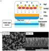
(a) Optical D-type fiber immunosensor functionalized with poly dimethyl diallyl ammonium chloride (PDDA) and poly(sodium-p-styrenesulfonate) (PSS) to immobilize goat anti-human IgG (GaHIgG) for detecting human IgG (HIgG) (adapted with permission from [79], Copyright Elsevier 2020). (b) Top and side SEM images of SiO2/ZnO nanowires (adapted from [88] with permission from The Royal Society of Chemistry).
Figure 3
Figure 3
(a) Schematic representation of a magneto-actuated biosensor where commercial magnetic particles were modified with an anti-CD3 antibody to isolate CD4+ cells from monocytes and macrophages. Biotinylated anti-CD4 antibodies labeled with streptavidin-horseradish peroxidase (HRP) were used to label the isolated CD4+ cells. The amperometric measurement was mediated by H2O2 and hydroquinone (adapted with permission from [93], Copyright Elsevier 2015). (b) Example of a cytotoxic T cell adhered on top of a transistor gate with a dimension of 12 × 5 mm2 (adapted with permission from [99], Copyright Elsevier 2015). (c) Schematic representation of a label-free Surface plasmon resonance (SPR) biosensor to analyze tumor-specific CD8+ cells. The gold layer of the SPR sensor was coated with planar lipid bilayers to immobilize major histocompatibility complex with antigen-derived peptides (p/MHC) that bound with CD8+ cells (TCR, T cell receptor) (adapted with permission from [102], Copyright 2018 American Chemical Society). (d) Schematic representation of barcode nanowires of alternated Fe and Au multilayers to capture CD8+ cells and their secreted interferon-γ (IFN-γ). The iron segment was treated with 11-aminoundecanoic acid for binding anti-CD8, whereas thiolated anti-IFN-γ antibodies were conjugated to the Au segment (adapted with permission from [105], Copyright 2019 American Chemical Society).

References

    1. Omman R.A., Kini A.R. Leukocyte development, kinetics, and functions. In: Keohane E.M., Otto C.N., Walenga J.M., editors. Rodak’s Hematology: Clinical Principles and Applications. Saunders (Elsevier); Philadelphia, PA, USA: 2019. pp. 117–135.
    1. Cohn L., Hawrylowicz C., Ray A. Biology of Lymphocytes. In: Adkinson N.F., Bochner B.S., Burks A.W., Busse W.W., Holgate S.T., Lemanske R.F., O’Hehir R.E., editors. Middleton’s Allergy. 8th ed. Elsevier; London, UK: 2014. pp. 203–214.
    1. Alberts B., Johnson A., Lewis J., Raff M., Roberts K., Walter P. Molecular Biology of the Cell. 4th ed. Garland Science; New York, NY, USA: 2002. Lymphocytes and the Cellular Basis of Adaptive Immunity; pp. 125–152.
    1. Mollarasouli F., Kurbanoglu S., Ozkan S.A. The Role of Electrochemical Immunosensors in Clinical Analysis. Biosensors. 2019;9:86. doi: 10.3390/bios9030086. - DOI - PMC - PubMed
    1. Lim S.A., Ahmed M.U. Introduction to Immunosensors. In: Ahmed M.U., Zourob M., Tamiya E., editors. Detection Science. Royal Society of Chemistry; Cambridge, UK: 2019. pp. 1–20.

LinkOut - more resources