Review
. 2020 Oct 27;9(11):3457.
doi: 10.3390/jcm9113457.

Cytokines and Pathogenesis of Central Retinal Vein Occlusion

Affiliations
Review

Cytokines and Pathogenesis of Central Retinal Vein Occlusion

Hidetaka Noma et al. J Clin Med. .

Abstract

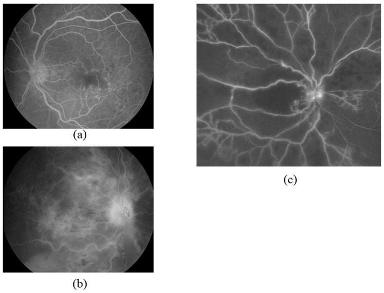
Central retinal vein occlusion (CRVO) causes macular edema and subsequent vision loss and is common in people with diseases such as arteriosclerosis and hypertension. Various treatments for CRVO-associated macular edema have been trialed, including laser photocoagulation, with unsatisfactory results. However, when the important pathogenic role of vascular endothelial growth factor (VEGF) in macular edema was identified, the treatment of CRVO was revolutionized by anti-VEGF therapy. However, despite the success of intraocular injection of anti-VEGF agents in many patients with CRVO, some patients continue to suffer from refractory or recurring edema. In addition, the expression of inflammatory cytokines increases over time, causing more severe inflammation and a condition that is increasingly resistant to anti-VEGF therapy. This indicates that the pathogenesis of macular edema in CRVO is more complex than originally thought and may involve factors or cytokines associated with inflammation and ischemia other than VEGF. CRVO is also associated with leukocyte abnormalities and a gradual reduction in retinal blood flow velocity, which increase the likelihood of it developing from the nonischemic type into the more severe ischemic type; in turn, this results in excessive VEGF expression and subsequent neovascular glaucoma. Here, we review the role of different factors and cytokines involved in CRVO pathogenesis and propose a mechanism that holds promise for the development of novel therapies.

Keywords: central retinal vein occlusion; cytokines; macular edema; neovascularization.


Conflict of interest statement

The authors declare no conflict of interest.

Figures

Figure 1
Figure 1
Images of central retinal vein occlusion (CRVO). (a) Color fundus photograph of nonischemic CRVO. (b) Fluorescein angiogram showing ischemic CRVO. (c) Mild retinal hypoxia in a case of nonischemic CRVO. (d) Severe retinal hypoxia in a case of ischemic CRVO. (e,f) Optical coherence tomography images of macular edema and serous retinal detachment in (e) a case of nonischemic CRVO (in which they can occur even if retinal hypoxia is mild) and (f) a case of ischemic CRVO (in which they are common).
Figure 2
Figure 2
Representative fluorescein angiograms indicating that the severity of ischemia correlates to the concentration of vascular endothelial growth factor (VEGF) in the aqueous humor in cases of retinal nonperfusion. Values shown are aqueous VEGF levels; they are typical of values observed in mild, moderate, and severe cases of ischemia. (a) Mild ischemia (41,3 pg/mL). (b) Moderate ischemia (327 pg/mL). (c) Severe ischemia (1572 pg/mL).
Figure 3
Figure 3
Mechanism of macular edema pathogenesis in central retinal vein occlusion (CRVO). CRVO causes ischemia and produces inflammation that is secondary to impaired retinal perfusion and hemorrhage. This is associated with an upregulation of VEGF and inflammatory cytokine expression, which disrupts the blood–retinal barrier (BRB) and ultimately results in macular edema. VEGF and PlGF activate VEGFR-1, leading to leukocyte recruitment and further increasing inflammatory cytokine expression. In addition, VEGF activates VEGFR-2, which increases vascular permeability and further enhances the expression of inflammatory cytokines such as MCP-1 and ICAM-1 via NF-κB. The resultant chemotaxis and adhesion of leukocytes to the vascular endothelium decreases blood flow velocity. Blood flow velocity also worsens with increasing retinal edema, which results in longer diffusion distances. Decreased blood flow velocity thus creates a positive feedback loop that further exacerbates ischemia. Treatment of edema also compensates for the relative ischemia and leads to less leakage into the tissue. The resulting phagocytosis of leaking material, blood, and ischemic tissue may be an important contributor to maintaining and worsening the inflammation. As a result, the pathological mechanism increases in complexity. This hypothesis predicts that inflammation gains influence in the pathogenesis of CRVO as it develops into a chronic condition.
Figure 4
Figure 4
Mechanism of neovascularization pathogenesis in central retinal vein occlusion (CRVO). CRVO is associated with elevated expression of inflammatory cytokines such as MCP-1 and ICAM-1, which leads to leukocyte abnormalities and a decrease in retinal blood flow velocity. The slowing of the blood flow creates a positive feedback loop that further exacerbates ischemia. In addition, the resultant leukocyte chemotaxis and adhesion enhance inflammation, creating a second positive feedback loop. As a result, the risk of CRVO developing from nonischemic to ischemic increases. Finally, hypoxia causes excessive overexpression of VEGF, which results in vitreous hemorrhage and neovascular glaucoma.

References

    1. Mohamed Q., McIntosh R.L., Saw S.M., Wong T.Y. Interventions for central retinal vein occlusion: An evidence-based systematic review. Ophthalmology. 2007;114:507–519. doi: 10.1016/j.ophtha.2006.11.011. - DOI - PubMed
    1. Brown D.M., Campochiaro P.A., Singh R.P., Li Z., Gray S., Saroj N., Rundle A.C., Rubio R.G., Murahashi W.Y. Ranibizumab for macular edema following central retinal vein occlusion: Six-month primary end point results of a phase III study. Ophthalmology. 2010;117:1124–1133. doi: 10.1016/j.ophtha.2010.02.022. - DOI - PubMed
    1. Noma H., Yasuda K., Shimura M. Cytokines and the Pathogenesis of Macular Edema in Branch Retinal Vein Occlusion. J. Ophthalmol. 2019;2:5185128. doi: 10.1155/2019/5185128. - DOI - PMC - PubMed
    1. Hayreh S.S., Zimmerman M.B., Podhajsky P. Incidence of various types of retinal vein occlusion and their recurrence and demographic characteristics. Am. J. Ophthalmol. 1994;117:429–441. doi: 10.1016/S0002-9394(14)70001-7. - DOI - PubMed
    1. Hayreh S.S. So-called “central retinal vein occlusion”. I. Pathogenesis, terminology, clinical features. Int. J. Ophthalmol. 1976;172:1. - PubMed

LinkOut - more resources