Review
. 2020 Oct 27;9(11):2365.
doi: 10.3390/cells9112365.

Neurofibromin Structure, Functions and Regulation

Affiliations
Review

Neurofibromin Structure, Functions and Regulation

Mohammed Bergoug et al. Cells. .

Abstract

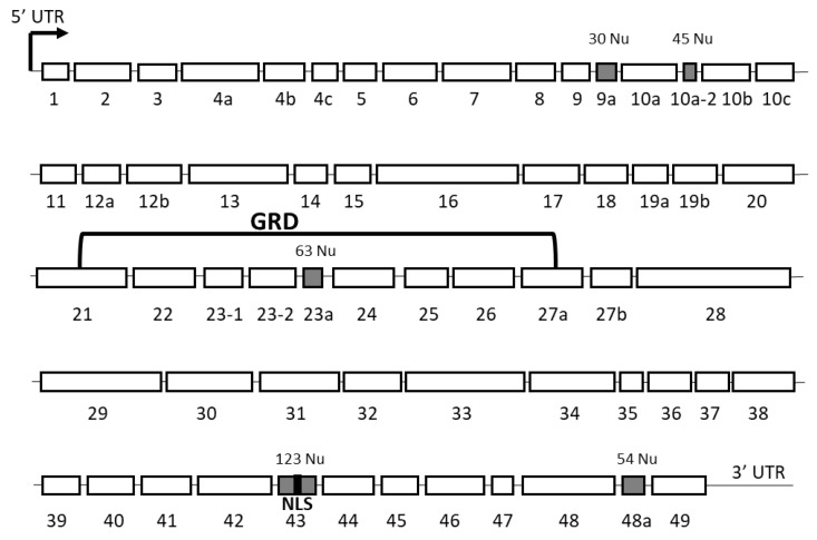
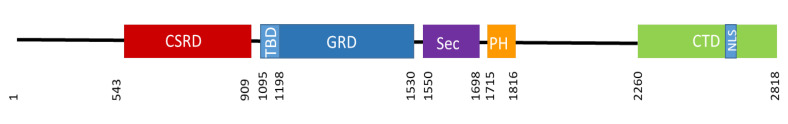
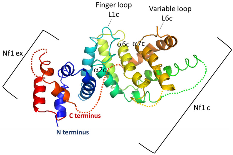
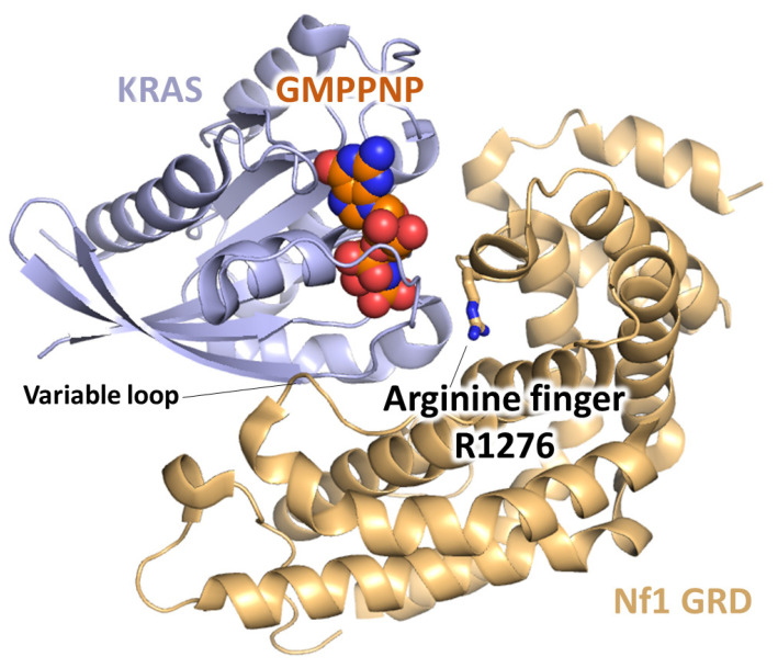
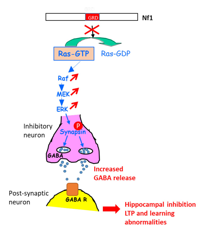
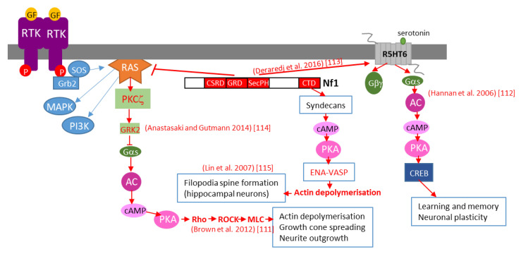
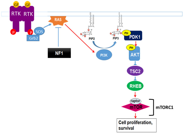
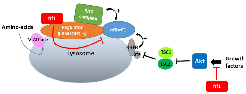
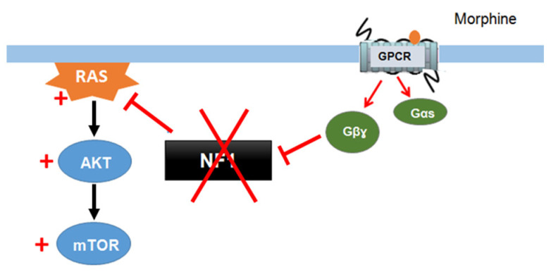
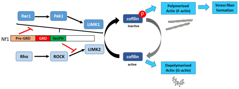
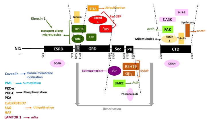
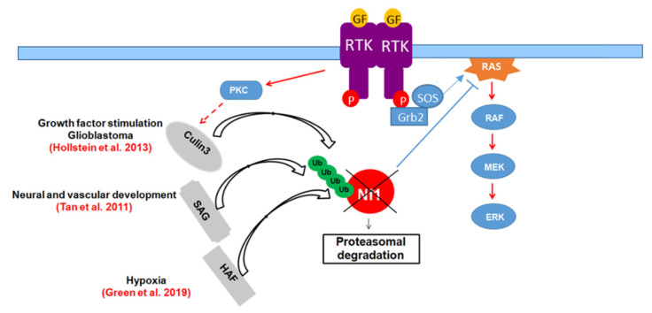
Neurofibromin is a large and multifunctional protein encoded by the tumor suppressor gene NF1, mutations of which cause the tumor predisposition syndrome neurofibromatosis type 1 (NF1). Over the last three decades, studies of neurofibromin structure, interacting partners, and functions have shown that it is involved in several cell signaling pathways, including the Ras/MAPK, Akt/mTOR, ROCK/LIMK/cofilin, and cAMP/PKA pathways, and regulates many fundamental cellular processes, such as proliferation and migration, cytoskeletal dynamics, neurite outgrowth, dendritic-spine density, and dopamine levels. The crystallographic structure has been resolved for two of its functional domains, GRD (GAP-related (GTPase-activating protein) domain) and SecPH, and its post-translational modifications studied, showing it to be localized to several cell compartments. These findings have been of particular interest in the identification of many therapeutic targets and in the proposal of various therapeutic strategies to treat the symptoms of NF1. In this review, we provide an overview of the literature on neurofibromin structure, function, interactions, and regulation and highlight the relationships between them.

Keywords: function; interactions; localization; neurofibromin; post-translational modifications; structure.


Conflict of interest statement

The authors declare no conflict of interest.

Figures

Figure 1
Figure 1
Schematic representation of the NF1 gene and its mRNA transcript; kb: kilo bases
Figure 2
Figure 2
Schematic representation of the exons of the full-length transcript of NF1 according to the old nomenclature. The alternatively spliced exons are indicated in grey with their number of nucleotides. The nuclear localization signal (NLS) in exon 43 (in black) and the GAP-related domain (GRD) are indicated; Nu: nucleotides.
Figure 3
Figure 3
Schematic representation of neurofibromin domains. CSRD (cysteine- and serine-rich domain) in red, TBD (tubulin-binding domain) in light blue, GRD (GAP-related domain) in blue, Sec (Sec14 homologous domain) in purple, PH (pleckstrin homologous domain) in orange, CTD (C-terminal domain) in green, NLS (nuclear localization signal) in blue. The amino acid number is indicated below.
Figure 4
Figure 4
Structure of neurofibromin-GRD domain (NF1-333) in a ribbon representation. The central domain (NF1c) is shown in light blue, green, yellow, and brown and the extra domain (NF1ex) in dark blue and red. Regions that are not visible in the neurofibromin-GRD model were complemented by the corresponding segments derived from the GAP-334 model and are shown as dotted lines. Helices α6c and α7c, forming the bottom of the Ras-binding groove, are indicated. The variable loop (L6c) and α2c helix, involved in the interaction with Ras, and finger loop (L1c), which provides an Arg residue (R1276) to the active site of Ras to stabilize the transition state of the GTPase reaction, are also indicated.
Figure 5
Figure 5
Structure of a KRas-neurofibromin GRD complex in a ribbon representation. The arginine finger (R1276) and variable loop are shown.
Figure 6
Figure 6
Structure of the SecPH domain: (A) Ribbon representation of the SecPH domain of human neurofibromin [50]. (B) Ribbon representation of neurofibromin-SecPH superimposed over the open Sec conformation (derived from the structure of Sec14p) [51].
Figure 7
Figure 7
Proposed functional mechanism of the neurofibromin-SecPH module. Hypothetical mechanism of how conformational changes in the neurofibromin-PH domain upon binding of ligand A may regulate access to ligand B for the neurofibromin-Sec lipid-binding cage.
Figure 8
Figure 8
Schematic representation explaining the increase in GABA secretion upon the activation of ERK in NF1-deficient inhibitory neurons. GABA R: GABA receptor.
Figure 9
Figure 9
Schematic representation of various mechanisms of neurofibromin-mediated cAMP regulation. GF: growth factor, RTK: tyrosine kinase receptor, AC: adenylate cyclase, CREB: cAMP response element-binding protein, MAPK: mitogen-activated protein kinase, PI3K: phosphoinositide 3-kinase, SOS: son of sevenless, Grb2: growth factor receptor-bound protein 2.
Figure 10
Figure 10
Neurofibromin regulation of the growth factor-controlled Ras/PI3K signaling pathway. PIP2: phosphatidylinositol-4,5-bisphosphate, PIP3: phosphatidylinositol 3,4,5 trisphosphate, PDK1: phosphoinositide-dependent kinase-1, PH: pleckstrin-homologous domain, TSC2: TSC complex subunit 2, RHEB: Ras homolog enriched in brain.
Figure 11
Figure 11
Neurofibromin binds to LAMTOR1 and inhibits mTORC1 signaling.
Figure 12
Figure 12
Neurofibromin mediates cross talk between GPCR (opioid receptors) and Ras/AKT signaling [132].
Figure 13
Figure 13
Schematic representation of the mechanisms for neurofibromin-mediated regulation of the actin cytoskeleton [136,137].
Figure 14
Figure 14
Neurofibromin interacting partners. Neurofibromin domains are indicated as black boxes and their limits are written below. Interacting partners of each domain are pictured between the dashed lines delimiting the domains. Each type of function is represented by a color: Ras-GTP in red, cAMP in brown, actin dynamics in green, microtubules in yellow, transport along microtubules in dark green, spinogenesis in purple, ubiquitination in orange, sumoylation in blue, phosphorylation in black, plasma membrane localization in dark blue, and unknown function in pink. Regions necessary for neurofibromin dimerization are indicated in grey. Partners interacting with unknown domains of neurofibromin or being part of the same complex are not pictured but their name is indicated on the left side of the figure.
Figure 15
Figure 15
Schematic representation of the mechanisms for neurofibromin-mediated inhibition of CRMP2 phosphorylation and inactivation [59,145].
Figure 16
Figure 16
Regulation of neurofibromin stability by the ubiquitin-proteasome pathway.

References

    1. Viskochil D., Buchberg A.M., Xu G., Cawthon R.M., Stevens J., Wolff R.K., Culver M., Carey J.C., Copeland N.G., Jenkins N.A., et al. Deletions and a translocation interrupt a cloned gene at the neurofibromatosis type 1 locus. Cell. 1990;62:187–192. doi: 10.1016/0092-8674(90)90252-A. - DOI - PubMed
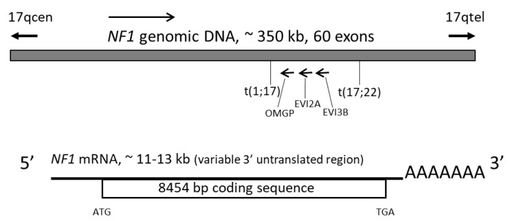
    1. Wallace M.R., Andersen L.B., Fountain J.W., Odeh H.M., Viskochil D., Marchuk D.A., O’Connell P., White R., Collins F.S. A chromosome jump crosses a translocation breakpoint in the von Recklinghausen neurofibromatosis region. Genes Chromosomes Cancer. 1990;2:271–277. doi: 10.1002/gcc.2870020404. - DOI - PubMed
    1. Henkemeyer M., Rossi D.J., Holmyard D.P., Puri M.C., Mbamalu G., Harpal K., Shih T.S., Jacks T., Pawson T. Vascular system defects and neuronal apoptosis in mice lacking ras GTPase-activating protein. Nature. 1995;377:695–701. doi: 10.1038/377695a0. - DOI - PubMed
    1. Koczkowska M., Chen Y., Callens T., Gomes A., Sharp A., Johnson S., Hsiao M.C., Chen Z., Balasubramanian M., Barnett C.P., et al. Genotype-Phenotype Correlation in NF1: Evidence for a More Severe Phenotype Associated with Missense Mutations Affecting NF1 Codons 844–848. Am. J. Hum. Genet. 2018;102:69–87. doi: 10.1016/j.ajhg.2017.12.001. - DOI - PMC - PubMed
    1. Gutmann D.H., Ferner R.E., Listernick R.H., Korf B.R., Wolters P.L., Johnson K.J. Neurofibromatosis type 1. Nat. Rev. Dis. Prim. 2017;3:17004. doi: 10.1038/nrdp.2017.4. - DOI - PubMed

Publication types