Review
. 2020 Oct 29;20(1):257.
doi: 10.1186/s12893-020-00921-6.

Living donor liver transplantation for idiopathic portal hypertension with extrahepatic portal vein stenosis and splenic artery aneurysms: a case report and review of the literature

Affiliations
Review

Living donor liver transplantation for idiopathic portal hypertension with extrahepatic portal vein stenosis and splenic artery aneurysms: a case report and review of the literature

Shigeyuki Kawachi et al. BMC Surg. .

Abstract

Background: Idiopathic portal hypertension (IPH) generally has a good prognosis and rarely results in liver transplantation. Furthermore, there are few reports of living donor liver transplantation (LDLT) for IPH with extrahepatic portal vein stenosis.

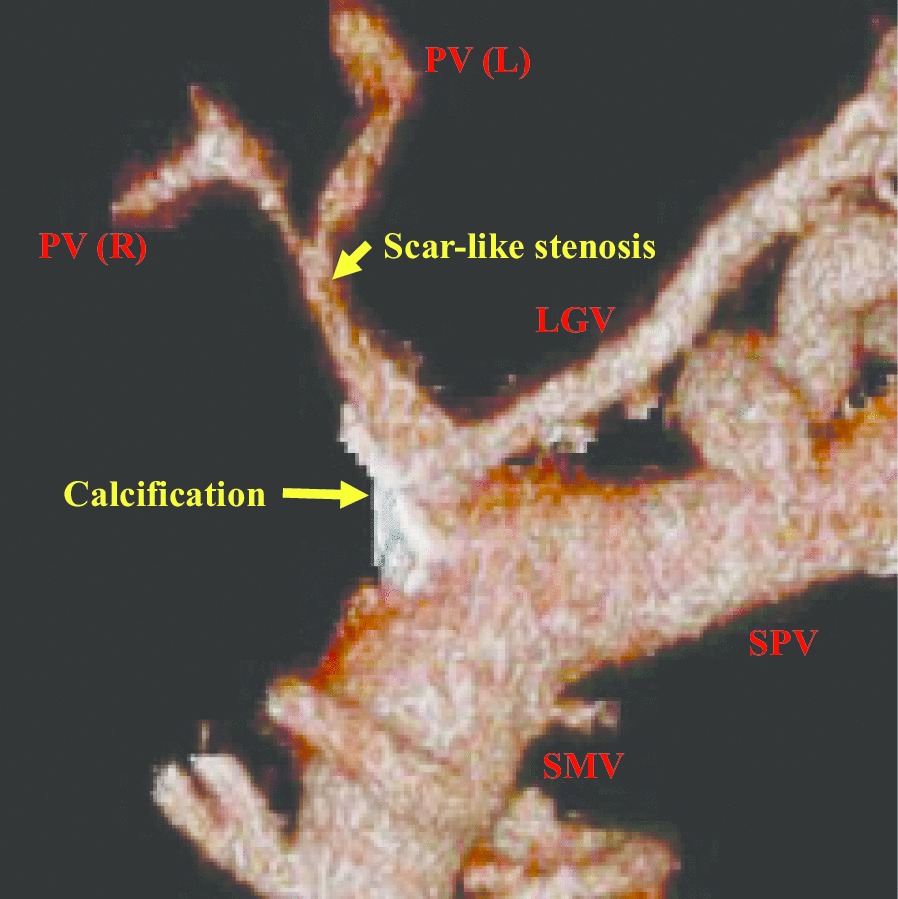
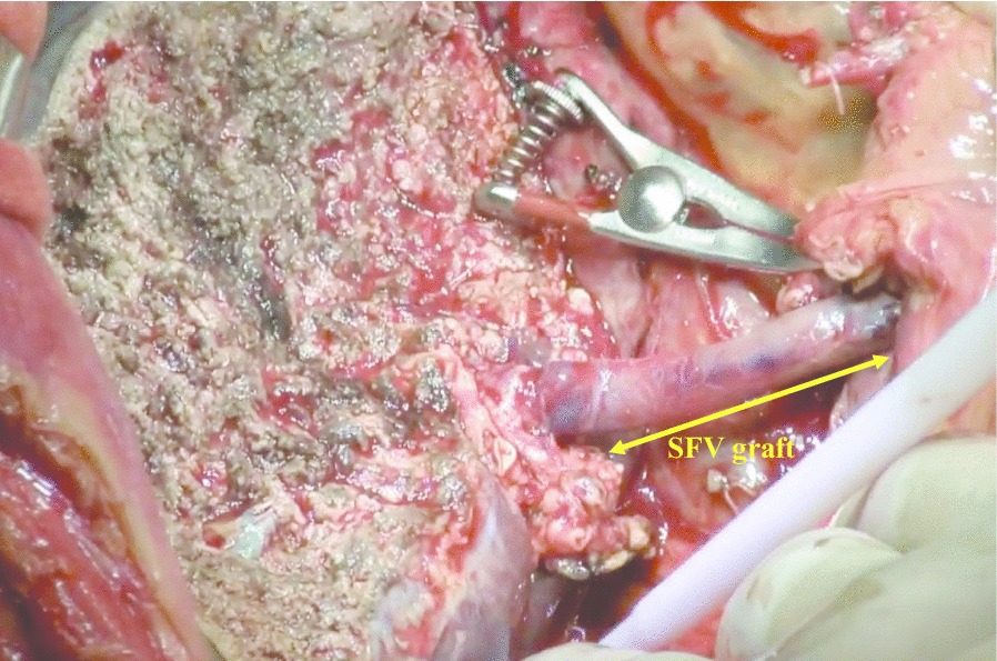
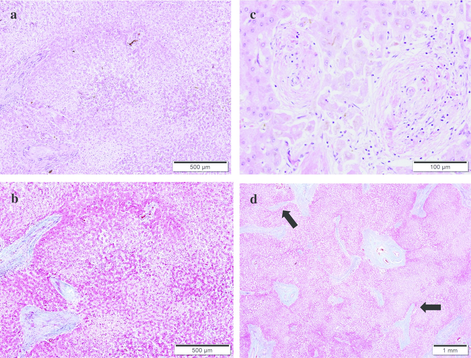
Case presentation: We report the case of a 51-year-old female patient diagnosed with IPH more than 20 years ago. She suffered severe jaundice, massive ascites, and encephalopathy at the time of her visit to our hospital. The patient's extrahepatic portal vein showed a scar-like stenosis, and the portal flow was completely hepatofugal. Collateral circulation such as the splenorenal shunt was well developed, and multiple splenic artery aneurysms up to 2 cm were observed in the splenic hilum. Her Model for End-Stage Liver Disease score increased to over 40 because of renal dysfunction, requiring temporary dialysis. We performed LDLT using her husband's right lobe graft and splenectomy. The extrahepatic stenotic portal vein was completely resected, and the superficial femoral vein (SFV) graft collected from the recipient's right leg was used for portal reconstruction as an interposition graft. Although the clinical course after LDLT had many complications, the patient was discharged on postoperative day 113 and has been fine for 2 years after LDLT. Histopathologically, the explanted liver had obliterative portal venopathy, nodular regenerative hyperplasia, and incomplete septal cirrhosis.

Conclusion: This case showed that severe IPH is occasionally associated with extrahepatic portal vein stenosis and can be treated with LDLT with portal vein reconstruction using an interposition graft. It was also suggested that the SFV is a useful choice for the interposition graft.

Keywords: Extrahepatic portal vein stenosis; Idiopathic portal hypertension; Living donor liver transplantation; Splenic artery aneurysms; Superficial femoral vein graft.


Conflict of interest statement

The authors declare that they have no competing interests.

Figures

Fig. 1
Fig. 1
A 3D preoperative portal venous image. The extrahepatic portal vein showed a scar-like stenosis, and collateral circulation was well developed. Dense calcification was observed at the site of the splenomesenteric junction
Fig. 2
Fig. 2
The arterial phase of abdominal dynamic CT. The arterial phase of abdominal dynamic CT reveals the multiple splenic artery aneurysms up to 2 cm in size in the splenic hilum
Fig. 3
Fig. 3
Intraoperative photo after portal vein reconstruction. Portal reconstruction was performed using the interposition SFV graft between the right lobe graft portal vein stump and the splenomesenteric junction
Fig. 4
Fig. 4
Histologic feature of the explanted liver. a Nodular regenerative hyperplasia (NRH) was noted in the liver explants. Parenchymal nodularity created by areas of hypertrophic hepatocytes in periportal areas alternating with areas of cord atrophy without fibrosis (H&E stain). b Same field of view as A by Masson trichrome stain. c Obliterative portal venopathy was seen in this portal tract, in which there is a diminished-caliber and almost sclerotic portal vein (H&E stain). d Incomplete septal cirrhosis was observed in this slide. Here, fibrous bands (arrows) extend from the portal tract and end blindly in the liver parenchyma (Masson trichrome stain)
Fig. 5
Fig. 5
Portal venous image 2 years after LDLT. Portal venous image was constructed two years after LDLT. The interposition SFV graft was well patent and no anastomotic stenosis was observed in both, the distal and proximal anastomotic sites

Similar articles

Cited by

References

    1. Tanaka T, Sugawara Y, Kokudo N. The current clinical aspects of idiopathic portal hypertension. Intractable Rare Dis Res. 2013;2:94–97. - PMC - PubMed
    1. Khanna R, Sarin SK. Idiopathic portal hypertension and extrahepatic portal venous obstruction. Hepatol Int. 2018;12:S148–S167. doi: 10.1007/s12072-018-9844-3. - DOI - PubMed
    1. Krasinskas AM, Eghtesad B, Kamath PS, Demetris AJ, Abraham SC. Liver transplantation for severe intrahepatic noncirrhotic portal hypertension. Liver Transplant. 2005;11:627–634. doi: 10.1002/lt.20431. - DOI - PubMed
    1. Meijer B, Simsek M, Blokzijl H, Man RA, Coenraad MJ, Dijkstra G, et al. Nodular regenerative hyperplasia rarely leads to liver transplantation: a 20-year cohort study in all Dutch liver transplant units. United Eur Gastroenterol J. 2017;5:658–667. doi: 10.1177/2050640616680550. - DOI - PMC - PubMed
    1. Inokuma T, Eguchi S, Tomonaga T, Miyazaki K, Hamasaki K, Tokai H, et al. Acute Deterioration of idiopathic portal hypertension requiring living donor liver transplantation: a case report. Dig Dis Sci. 2009;54:1597–1601. doi: 10.1007/s10620-008-0504-7. - DOI - PubMed

MeSH terms